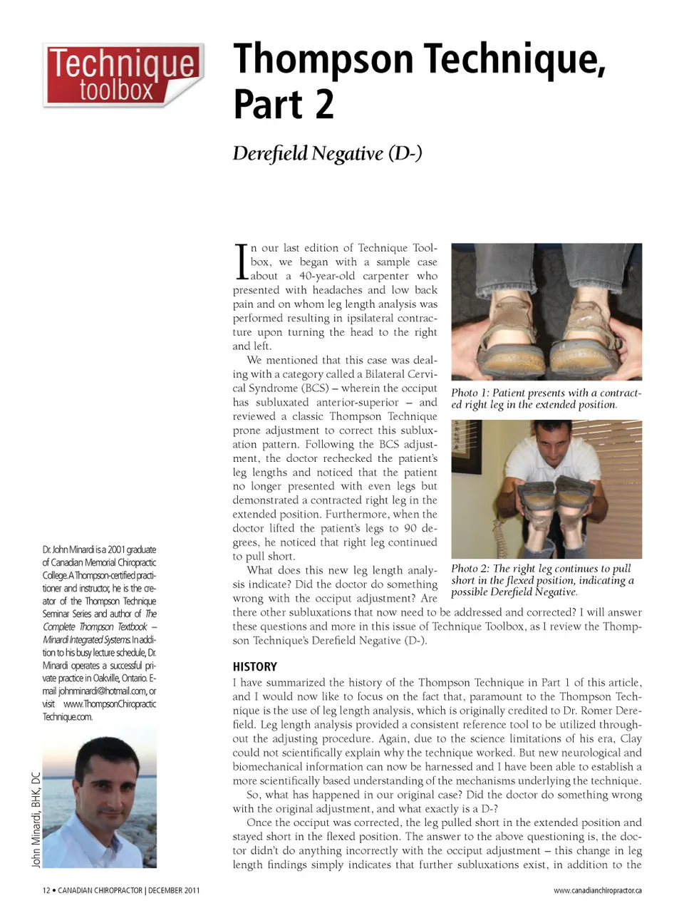
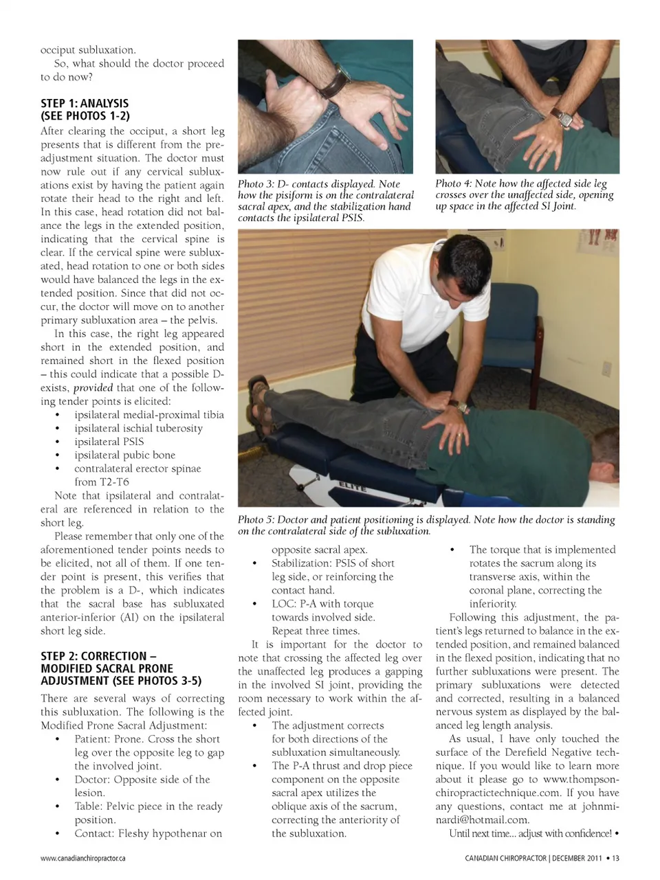
occiput subluxation. So, what should the doctor proceed to do now? stEP 1: analysis (sEE Photos 1-2) After clearing the occiput, a short leg presents that is different from the pre-adjustment situation. The doctor must now rule out if any cervical sublux -ations exist by having the patient again rotate their head to the right and left. In this case, head rotation did not bal -ance the legs in the extended position, indicating that the cervical spine is clear. If the cervical spine were sublux -ated, head rotation to one or both sides would have balanced the legs in the ex -tended position. Since that did not oc -cur, the doctor will move on to another primary subluxation area – the pelvis. In this case, the right leg appeared short in the extended position, and remained short in the flexed position – this could indicate that a possible D-exists, provided that one of the follow -ing tender points is elicited: • ipsilateral medial-proximal tibia • ipsilateral ischial tuberosity • ipsilateral PSIS • ipsilateral pubic bone • contralateral erector spinae from T2-T6 Note that ipsilateral and contralat -eral are referenced in relation to the short leg. Please remember that only one of the aforementioned tender points needs to be elicited, not all of them. If one ten -der point is present, this verifies that the problem is a D-, which indicates that the sacral base has subluxated anterior-inferior (AI) on the ipsilateral short leg side. stEP 2: CorrECtion – moDifiED saCral PronE aDJustmEnt (sEE Photos 3-5) There are several ways of correcting this subluxation. The following is the Modified Prone Sacral Adjustment: • Patient: Prone. Cross the short leg over the opposite leg to gap the involved joint. • Doctor: Opposite side of the lesion. • Table: Pelvic piece in the ready position. • Contact: Fleshy hypothenar on www.canadianchiropractor.ca Photo 3: D-contacts displayed. Note how the pisiform is on the contralateral sacral apex, and the stabilization hand contacts the ipsilateral PSIS. Photo 4: Note how the affected side leg crosses over the unaffected side, opening up space in the affected SI Joint. Photo 5: Doctor and patient positioning is displayed. Note how the doctor is standing on the contralateral side of the subluxation. opposite sacral apex. Stabilization: PSIS of short leg side, or reinforcing the contact hand. • LOC: P-A with torque towards involved side. Repeat three times. It is important for the doctor to note that crossing the affected leg over the unaffected leg produces a gapping in the involved SI joint, providing the room necessary to work within the af -fected joint. • The adjustment corrects for both directions of the subluxation simultaneously. • The P-A thrust and drop piece component on the opposite sacral apex utilizes the oblique axis of the sacrum, correcting the anteriority of the subluxation. • The torque that is implemented rotates the sacrum along its transverse axis, within the coronal plane, correcting the inferiority. Following this adjustment, the pa -tient’s legs returned to balance in the ex -tended position, and remained balanced in the flexed position, indicating that no further subluxations were present. The primary subluxations were detected and corrected, resulting in a balanced nervous system as displayed by the bal -anced leg length analysis. As usual, I have only touched the surface of the Derefield Negative tech -nique. If you would like to learn more about it please go to www.thompson -chiropractictechnique.com. If you have any questions, contact me at johnmi [email protected]. Until next time... adjust with confidence! • CANADIAN CHIROpRACTOR | DECEMBER 2011 • 13 •
Chiropractic + Naturopathic Doctor December 2011: Page 13