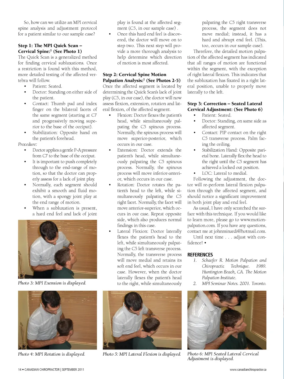
So, how can we utilize an MPI cervical spine analysis and adjustment protocol for a patient similar to our sample case? Step 1: The MPI Quick Scan – Cervical Spine: 2 (See Photo 1) The Quick Scan is a generalized method for finding cervical subluxations. Once a restriction is found with this method, more detailed testing of the affected ver-tebra will follow. • Patient: Seated. • Doctor: Standing on either side of the patient. • Contact: Thumb pad and index finger on the bilateral facets of the same segment (starting at C7 and progressively moving supe-rior to the base of the occiput). • Stabilization: Opposite hand on the patient’s forehead. Procedure: • Doctor applies a gentle P-A pressure from C7 to the base of the occiput. • It is important to push completely through to the end-range of mo-tion, so that the doctor can prop-erly assess for a lack of joint play. • Normally, each segment should exhibit a smooth and fluid mo-tion, with a spongy joint play at the end range of motion. • When a subluxation is present, a hard end feel and lack of joint • play is found at the affected seg-ment (C5, in our sample case) . Once this hard end feel is discov-ered, the doctor will move on to step two. This next step will pro-vide a more thorough analysis to help determine which direction of motion is most affected. Photo 3: MPI Exension is displayed. Step 2: Cervical Spine Motion Palpation Analysis: 2 (See Photos 2-5) Once the affected segment is located by determining the Quick Scan’s lack of joint play (C5, in our case), the doctor will now assess flexion, extension, rotation and lat-eral flexion, of the affected segment. • Flexion: Doctor flexes the patient’s head, while simultaneously pal-pating the C5 spinous process. Normally, the spinous process will move superior-posterior, which occurs in our case. • Extension: Doctor extends the patient’s head, while simultane-ously palpating the C5 spinous process. Normally, the spinous process will move inferior-anteri-or, which occurs in our case. • Rotation: Doctor rotates the pa-tient’s head to the left, while si-multaneously palpating the C5 right facet. Normally, the facet will move anterior-superior, which oc-curs in our case. Repeat opposite side, which also produces normal findings in this case. • Lateral Flexion: Doctor laterally flexes the patient’s head to the left, while simultaneously palpat-ing the C5 left transverse process. Normally, the transverse process will move medial and retains its soft end feel, which occurs in our case. However, when the doctor laterally flexes the patient’s head to the right, while simultaneously palpating the C5 right transverse process, the segment does not move medial; instead, it has a hard and abrupt end feel. (This, too, occurs in our sample case). Therefore, the detailed motion palpa-tion of the affected segment has indicated that all ranges of motion are functional within the segment, with the exception of right lateral flexion. This indicates that the subluxation has fixated in a right lat-eral position, unable to properly move laterally to the left. Step 3: Correction – Seated Lateral Cervical Adjustment: (See Photo 6) • Patient: Seated. • Doctor: Standing, on same side as affected segment. • Contact: PIP contact on the right C5 transverse process. Palm fac-ing the ceiling. • Stabilization Hand: Opposite pari-etal bone. Laterally flex the head to the right until the C5 segment has achieved a locked out position. • LOC: Lateral to medial. Following the adjustment, the doc-tor will re-perform lateral flexion palpa-tion through the affected segment, and should notice a significant improvement in both joint play and end feel. As usual, I have only scratched the sur-face with this technique. If you would like to learn more, please go to www.motion-palpation.com. If you have any questions, contact me at [email protected]. Until next time . . . adjust with con-fidence! • REFERENCES 1. Schaefer R. Motion Palpation and Chiropractic Technique. 1989. Huntington Beach, CA. The Motion Palpation Institute. 2. MPI Seminar Notes. 2001. Toronto. Photo 4: MPI Rotation is displayed. 14 • Canadian ChiropraCtor | September 2011 Photo 5: MPI Lateral Flexion is displayed. Photo 6: MPI Seated Lateral Cervical Adjustment is displayed. www.canadianchiropractor.ca
Chiropractic + Naturopathic Doctor September 2011: Page 14