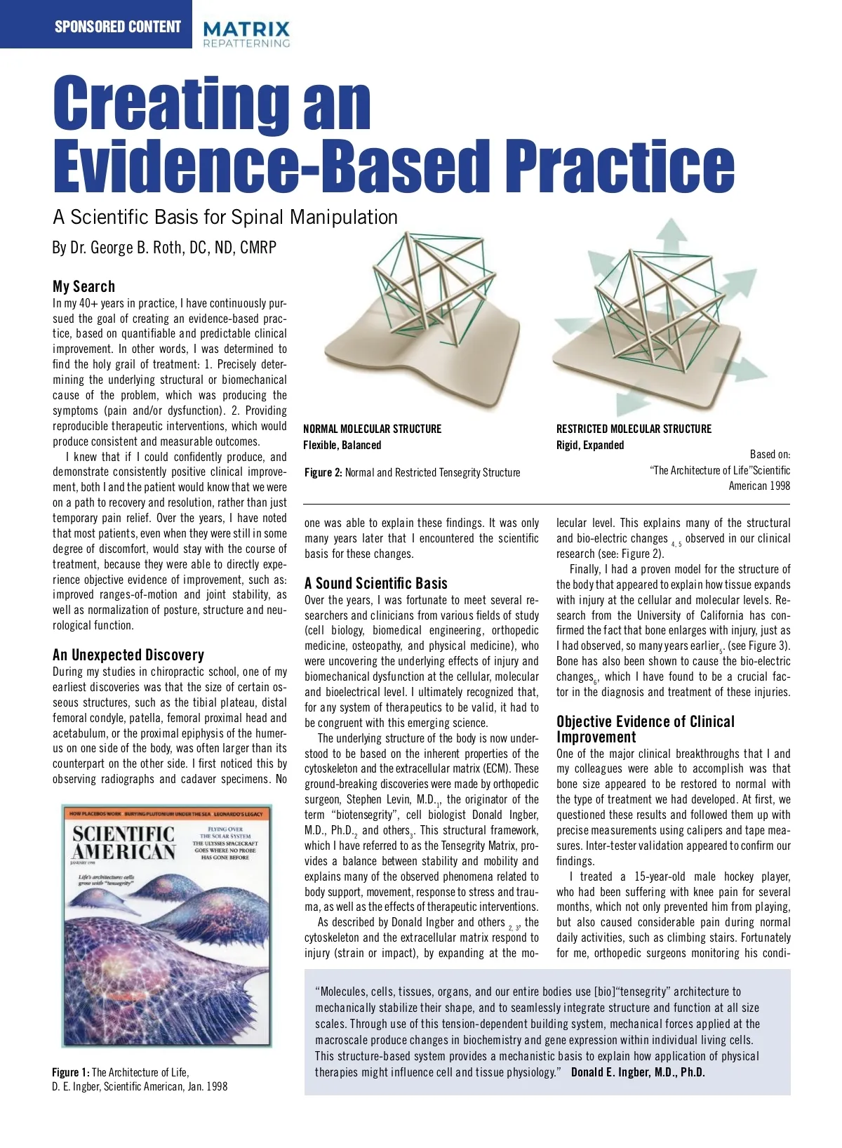
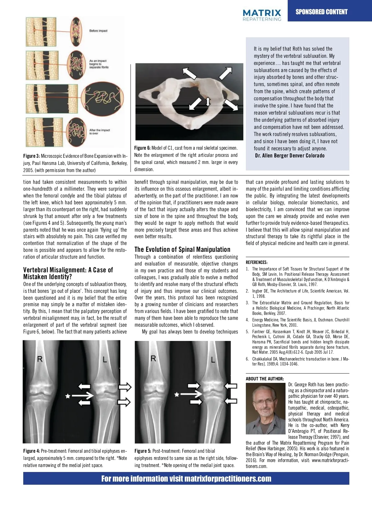
SPONSORED CONTENT SPONSORED CONTENT SPONSORED CONTENT R L to nd ed es ed. ay an ed of 1, ve ay re tly, if hat he ld re ve to sis us a ea nal he is ro-ng al-ny ng ns ve I have been a chiropractor for 2 over 40 years, in every these differences. At the time, these facts were the canal, which measured mm. larger that I realized how profound this omission of was. have been gratified to spinal note that many of them jury, Paul Hansma Lab, University California, Berkeley, but early the in my career, I became convinced that filed away, and it was only many years later that The more I investigated this important aspect have been able to reproduce same measur-dimension. 2005. (with permission from the author) I I was not achieving the kinds of results prom-these early observations came to be viewed in a of human anatomy and the common injuries, able outcomes, which observed. ised when I attended school. I also witnessed new light, based on my clinical research. which can to life-altering has always been to find measurable tion often had lead taken consistent outcomes, measurements My to goal within that can provide profound and lasting solutions to benefit through spinal manipulation, may be due to many of techniques my colleagues disheartened By carefully examining a better quality skel-C the more I recognized how important it was for evidence to support any of the we, becoming as one‐hundredth of a millimeter. They were surprised its or many of the painful and limiting conditions afflicting influence on this osseous enlargement, albeit in-failing practice. They had come into this etal model, which is cast from a real skeleton, me to incorporate a rational approach to the chiropractors, provide. I have in often wondered when the area. femoral condyle and the tibial plateau of be public. integrating the latest developments advertently, on part of the practitioner. I am you now can the profession high expectations and a sincere verify many By of these same discrepan-treatment of this how things might different if with our the profession to help their fellow humans, but the aware cies for yourself. Besides the examples of the the left knee, which had approximately mm. and in cellular biology, molecular biomechanics, and of desire the opinion if devel-practitioners were made By applying the principle of been identifying were to 5 embrace integrate the that, latest of treatments they were taught did shape not femur, and humerus mentioned earlier, and normalizing the of the cranial larger than its structure counterpart on the right, had suddenly opments in cell biology, bioelectricity, I am convinced that we can improve of types the molecular fact that biomechan-injury actually alters the and tibia live up to these expectations. a close inspection the we spine can be provide very re-and evolve even 4, bones, as with other areas of the body, have ics, biomedical engineering and bio-electricity shrunk by that amount after we only a few treatments upon the of care already size of bone in the spine and throughout the body, Like many of you, I pursued a long search vealing. Note the differences in the size (width, D witnessed a Figures remarkable degree of success in the 5 young . These man’s emerging they disciplines are crucial to to our (see 4 and 5). Subsequently, further to provide truly evidence‐based therapeutics. would be eager apply methods that would for additional modalities to improve my results, depth and height of the articular processes) at helping individuals recover from many dev-understanding of the effects of injury at the most parents noted that he was once again ‘flying up’ the more I believe that this the will allow Figure spinal 1 manipulation and precisely target these areas and thus achieve validate my outcomes and give me the confi-various levels throughout spine. astating neurological consequences, including fundamental level of the human body. It is my stairs with absolutely no pain. verified my scientific structural to in take rightful place in the even better results. dence to be able to find and resolve my patient’s demonstrates these therapy differences the its size of cognitive, visual, auditory, vestibular and This neu-case belief that these advances would sup-conditions. In this search, of medicine the atlas. and I contend contention that Several normalization of the shape of the field processes of physical health care in general. romuscular conditions. independent port much of what we already provide and help I was blessed to meet the articular several researchers clinicians from other that on palpation, these areas of enlargement Figure 2: Microscopic Eviden 3, allow 4 bone is possible and outcomes. appears to for the resto-The of and Spinal Manipulation researchers have verified these us evolve even further as Evolution a truly science-based Expansion with Injury, Paul fields (cell biology, biomedical engineering, may be easily mistaken for a relative rotation or University of California, Berk profession. I sense that there is a growing Dr. Norman Doidge, MD, who is on faculty ration of articular structure and function. Through a combination of relentless questioning orthopedic medicine, osteopathy and physical translation of the vertebral segment (see: Sub-permission from the author in evaluation our profession to see at Columbia University and the University of desire among many and of measurable, objective changes REFERENCES: medicine). were making amazing discov-luxation: A Case of Mistaken Identity, below). role They in the health Toronto, is a world-renowned expert in the chiropractic take its rightful 1. The Importance of Soft Tissues for Structural Support of the in eries my own practice and those effects of my and Vertebral Misalignment: A Case of the underlying of students injury Recent evidence from the of Cal-Therapy: male Assessment hockey player, who h in regarding the field of physical field of brain injury and neuroplasticity. After care industry, as leaders Body, SM Levin, In: University Positional Release colleagues, I was gradually able to evolve a method Mistaken Identity? and biomechanical dysfunction at the cellular, ifornia, revealed by the of powerful Atomic Dysfunction, Force from knee pain & Treatment Musculoskeletal K D’Ambrogio & for severa medicine. The way ahead benefits not only our hearing about our approach, he conducted an bio-electrical even the molecular level. I effects microscope, GB under direction St. of Louis, physicist to the identify and and resolve many of the structural One of the underlying of subluxation theory, Roth, the Mosby‐Elsevier, 1997. prevented him from playing profession but also countless individuals in-depth investigation, which concepts included obser-1 , has confirmed the for improve a system our of clinical therapeutics Paul Hansma and his caused considerable pain du 2. Ingber DE, team The Architecture of Life, Scientific American, Vol. of recognized injury and that thus outcomes. bones ‘go out of place’. This who has are long looking for real solutions for many vations is of that treatments, patient interviews with concept to be valid, it had be congruent with this activities, such as climbing presence of 1, certain 1998. protein structures within painful limiting conditions. numerous concussion and Over the years, this to protocol has been recognized been questioned and post-concussion it is my belief that the and entire emerging science. the bone that expand with an injury. These was only able to accomplis premise may simply be a matter of mistaken iden-by a growing number of clinicians and researchers 3. The Extracellular Matrix and Ground Regulation, Basis for bling manner. findings are a consistent with my clinical A obser-Holistic Biological Medicine, Pischinger, North Atlantic Fortunately f tity. By this, I mean that the palpatory perception of from various fields. I have been gratified to note that Books, Berkley, References: surgeons were monitoring th vations, which were first 2007. made over 40 years many of L, them have been able to reproduce the same vertebral misalignment fact, be the of H, Pechenik at the knee with a high de ago (see 4. Figure 2). Medicine, The Scientific Basis, JL Oschman. Energy Churchill 1. Fantner GE, Hassenkam T, may, Kindt in JH, Weaver JC, result Birkedal Cutroni JA, Cidade GA, Stucky GD, Morse Hansma PK, Sacrificial and hidden length which I observed. Livingstone, New York, 2001. due to an underlying genet measurable outcomes, enlargement of part of the DE, vertebral segment (see bonds dissipate energy as mineralized fibrils separate during bone fracture, Nat Mater. result, they took Restoration of GE, Bone Size T, Kindt JH, Weaver 5. Fantner Hassenkam JC, Birkedal H, consistent My goal has always been to develop techniques Figure 6, below). The fact that Jul many 2005 Aug;4(8):612-6. Epub 2005 17. patients achieve within one-hundredth of a and Joint Healing Pechenik L, Cutroni JA, Cidade GA, Stucky GD, Morse DE, 2. Doidge, N., The Brain’s Way of Healing, Penguin Books, New York, 2016. PK, clinical Sacrificial bonds and hidden were length dissipate all surprised when th One of the Hansma significant breakthroughs energy as mineralized fibrils separate bone 3. Tommerdahl, M, Dennis, RG, et al., Neurosensory Assessment of Concussion, Mil and the fracture, tibial plateau of th that my colleagues and I were able to ac-during Med. 2016 May;181(5 Suppl):45-50 Nat that Mater. 2005 size Aug;4(8):612‐6. Epub Jul 17. been approximately 5 m complish was bone appeared to be 2005 had 4. MacGuintie LA, Streaming and piezoelectric potentials in connective tissues, In: Chakkalakal Mechanoelectric transduction in bone. J Ma-on the right, had restored 6. to normal with DA, treatment. At first, we counterpart Blank M (ed) Electromagnetic fields: biological interactions and mechanisms. ter Res]. 1989;4: 1034‐1046. by that amount after only a f questioned these results and followed them up Advances in Chemistry Series 250. American Chemical Society, Washington DC, ch. Figure 3 and 4). Subsequentl with precise measurements using callipers and 8, pp 125-142, 1995. Figure 1: Model of C1, cast from a real parents noted that he was tape measures. validation appeared specimen. Note the enlargement 5. Chakkalakal DA, Mechanoelectric transduction in bone. J Mater skeletal Res]. 1989;4: 1034-1046. ABOUT Inter-tester THE AUTHOR: of the right articular process and the spinal up” the practic-stairs with absolutely to confirm our findings. Dr. George Roth has been canal, which measured 2 mm. larger in verified my contention that Several years ago, I treated ing a 15-year-old For more information: matrixforpractitioners.com as a chiropractor and a naturo-every dimension. experience… has taught me that vertebral subluxations are caused by the effects of injury absorbed by bones and other struc-tures, sometimes spinal, and often remote recently searched the Internet for scientific Startling Observations: The Evolution of Chiropractic with Injury! to support chiropractic. Unfortu-Bone Enlarges from the spine, which create patterns of Through a combination evidence of relentless question-most of the references emphasized While studying radiology throughout in chiropractic ing and evaluation of nately, measurable, objective compensation the body that the lack of scientific validation or measur-school, I noticed that the size of a structure on changes (biomechanical, structural, radio-involve the spine. I have found that the to support graphic, biochemical able and outcomes neurological) in my the basic tenets of one side of the body was often different than reason vertebral subluxations is that our profession. At best, there are a few studies its counterpart on the other side. For example, recur A Testable Hypothesis for a M own practice and those of my students and Resistance underlying patterns injury Figure 4, Post-treatment: Femoral and that demonstrate a modest femoral or humeral head, of or absorbed the Fracture colleagues, I was gradually able to evolve a advantage for chi-the proximal the tibial epiphyses restored to same size as A ropractic manipulation over some prescription tibial plateau and on one side was noticeably larger compensation have not been addressed. method to identify and resolve many of the the right side, following treatment. *Note medications for certain than the opposite side in the same individual. osseous (and other fascial) effects of injury and conditions, such as opening of the medial joint space. The work routinely resolves subluxations, back pain. However, My professors were unable to shed any light on thus improve our clinical outcomes. Over the the overall impression is and since I have been doing it, I have not that been chiropractic science years, this protocol has recognized by a is still relatively un-these findings. As an anatomy lab instructor at Figure 6: Model of C1, cast from a real specimen. found it subsequent necessary observations to adjust anyone. and that are skeletal only mini-the college and from structure of the cranium. It was only when I growing number of proven chiropractors, as chiropractors well as Note the enlargement of of the right articular process of and Allen Berger Denver Colorado mally accepted as part health care team. cadaverous Dr. specimens, I was able to confirm Figure 3: study Microscopic Evidence of Bone Expansion with In-researchers B embarked on a of osteopathic medicine clinicians and from various fields. I the patients, as well as participating in the training program himself. In his recent best-selling book, The Brain’s Way of Healing, he comments: “I view it as prudent to have a Matrix assessment A Subluxation: after a blow to the head… observing such cases has led me to hope By that Dr. soon, Matrix Repat-George Roth terning will be routinely applied in hospital emergency departments.” 2 Observations in a new light case of mistaken mystery identity? of the vertebral subluxation. My It is my belief that Roth has solved the I CC_Matrix_Sponsored_Content_DPS_Dec19_EJS.indd CC_Dec19_EJS.indd 18 2019-11-06 8:38 AM 2-3 Figure 4: Pre‐treatment: Femoral and tibial epiphyses en-larged, approximately 5 mm. compared to the right. *Note relative narrowing of the medial joint space. Figure 5: Post‐treatment: Femoral and tibial epiphyses restored to same size as the right side, follow-ing treatment. *Note opening of the medial joint space. pathic physician for over 40 years. He has taught at chiropractic, na-turopathic, medical, osteopathic, physical therapy and medical schools throughout North America. He is the co‐author, with Kerry D’Ambrogio PT, of Positional Re-lease Therapy (Elsevier, 1997), and the author of The Matrix Repatterning Program for Pain Relief (New Harbinger, 2005). His work is also featured in the Brain’s Way of Healing, by Dr. Norman Doidge (Penguin, 2016). For more information, visit: www.matrixforpracti-tioners.com. For more information visit matrixforpractitioners.com
Chiropractic + Naturopathic Doctor September/October 2022: Page 13