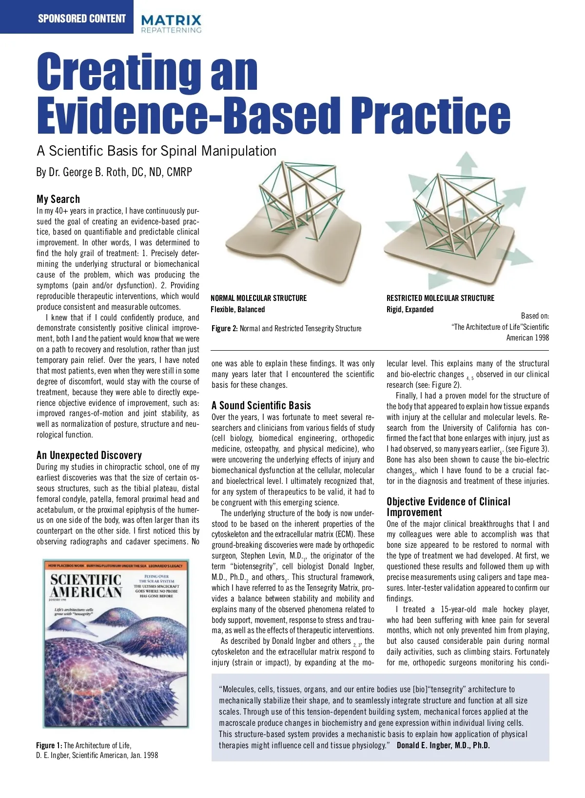
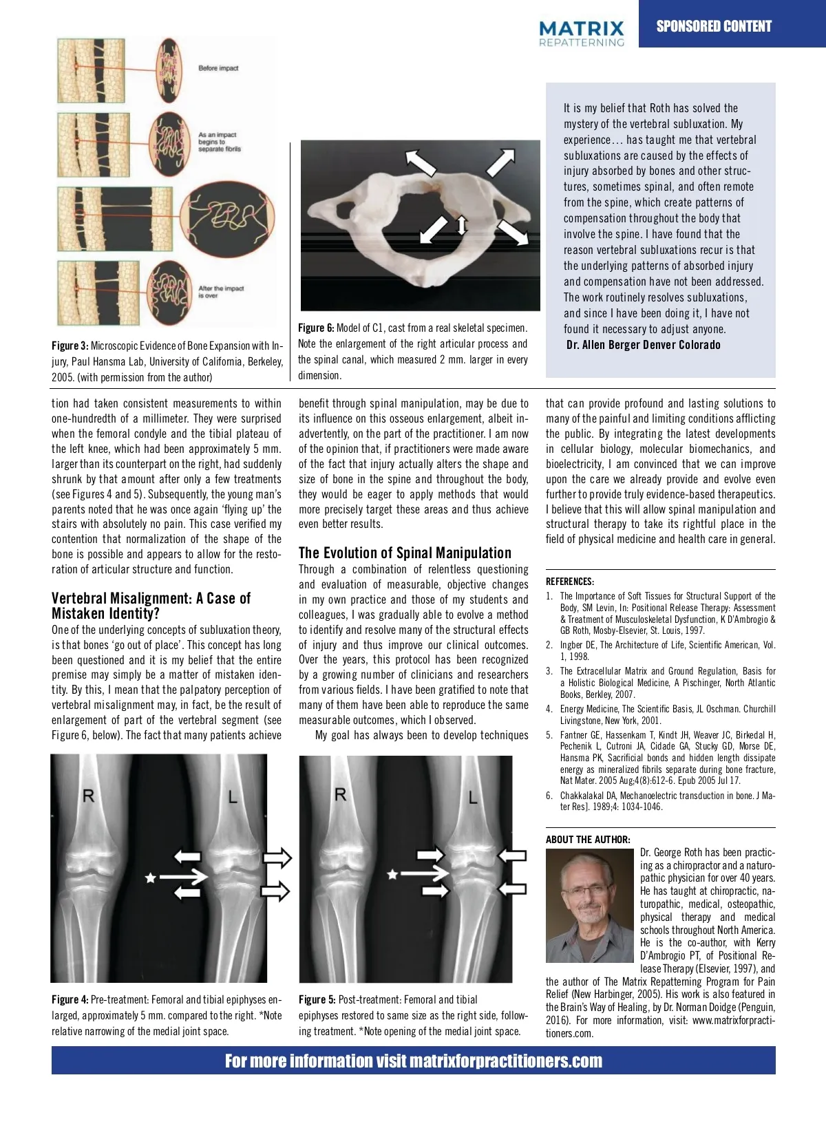
SPONSORED CONTENT CONTENTntly ation: A case of mistaken identity?bser . George Rothlux searched the Internet for scientific Startling Observations: Bone Enlarges with Injury! ence to support chiropractic. Unfortu-ly , most of the references emphasized While studying radiology in chiropractic lack of scientific validation or measur-tcomes school, I noticed that the size of a structure on to support the basic tenets of one side of the body was often different than fession. At best, there are a few studies its counterpart on the other side. For example, A Testable Hypothesis for a Molecular Origin of Figure 3, Pre-treatment: Femoral and monstrate a modest advantage for chi-ic the proximal femoral or humeral head, or the Fracture Resistance tibial epiphyses enlarged, approximately 5 manipulation over some prescription tibial plateau on one side was noticeably larger A My Search mm. compared to the right. *Note relative tions for certain conditions, such as than the opposite side in the same individual. narrowing of the medial joint space.NSORED In my 40+ years in practice, I have continuously pur-Before impact ain. However, the overall impression is My professors were unable to shed any light on sued the goal of creating an evidence‐based prac-iropractic science is still relatively un-and these findings. As an anatomy lab instructor at tice, based are on only quantifiable and predictable clinical that chiropractors mini-ccepted the college and from subsequent observations the shape of the bone is possible and appears improvement. In other I was determined as part of the health care team. words, of cadaverous specimens, to I was able to confirm B allow for the restoration of the joint space an find the for holy grail of treatment: 1. Precisely e been a chiropractor over 40 years, these differences. At deter-the time, these facts were As an impact biomechanical function, resulting in improve begins to y in my career, I became convinced that structural filed away, it was only many years later that mining the underlying or and biomechanical healing of the entire joint complex. separate fibrils ot achieving the kinds results prom-hen these early came to be viewed in a cause of of the problem, which was observations producing the I attended school. I also witnessed new light, based on my clinical research. Subluxation: symptoms (pain and/or dysfunction). 2. Providing f my colleagues becoming disheartened By carefully examining a better quality skel-C A Case of Mistaken Identity? reproducible therapeutic interventions, which would NORMAL MOLECULAR STRUCTURE RESTRICTED MOLECULAR STRUCTURE ng in practice. They had come into this etal model, which is cast from a real skeleton, The subluxation theory and the idea that bone consistent and measurable outcomes. on with high produce expectations and a sincere you can verify many of these same discrepan-Flexible, Balanced Rigid, Expanded “go out of place” has long been questione Based on: I knew that if I could produce, and to help their fellow humans, but the confidently cies for yourself. Besides the examples of the However, I believe that the entire premise ma “The of Life”Scientific f treatments demonstrate they were taught did not positive femur, tibia and improve-humerus mentioned earlier, consistently clinical Figure 2: Normal and Restricted Tensegrity Structure be Architecture a matter of mistaken identity. By this, I mea to these expectations.many close inspection spine can be very re-American 1998 of “misaligne that the palpatory impression ment, both I and the patient a would know that of we the were Dred_Content_DPS_Dec19_EJS.indd of you, I pursued a long search vealing. Note the differences in the size (width, vertebra” may be the result of enlargement on a path to recovery and resolution, rather than just itional modalities to improve my results, depth and height of the articular processes) at part of the vertebral segment (see Figure After the impact temporary relief. Over various the years, have noted the spine. my outcomes and give pain me the confi-o levels I throughout Figure 1 to explain these findings. It was only one was able many structural above). The of fact the that many patients achiev is over lecular level. This explains that most patients, even when they were still in some be able to find and resolve my patient’s demonstrates these differences many in the years size of later that I encountered the scientific and bio-electric changes observed our clinical benefit through in chiropractic adjustment ma 4, 5 degree of blessed discomfort, would stay with the course ons. In this search, I was to meet the articular processes of of the atlas. I for contend be due to its influence on the actual structu basis these changes. research (see: Figure 2). researchers and clinicians from other that able on palpation, these areas of enlargement Figure 2: Microscopic Evidence of Bone treatment, because they were to directly expe-of the osseous albeit Expansion with Injury, Paul Hansma Lab, Finally, I had a proven model for enlargement, the structure of inadvertent (cell biology, rience biomedical engineering, may be easily mistaken a relative rotation or University of California, Berkeley, 2005. (With on the part of the practitioner. In my opinion, objective evidence of improvement, such for as: the body that appeared to explain how tissue expands A Sound Scientific Basis edic medicine, osteopathy and physical translation of the vertebral segment (see: Sub-permission from the author) practitioners were made aware of the fact th improved ranges‐of‐motion and joint stability, as Over the years, I was fortunate to meet several re-with injury at the cellular and molecular levels. Re-of bone in th ne). They were making amazing discov-gar luxation: A Case of Mistaken Identity, below). injury alters the shape and size well as normalization of posture, structure and neu-ding the underlying effects of injury Recent evidence from the University of and Cal-clinicians search from the University of California has male hockey who had searchers from player, various fields of been study suffering spine and throughout the con-body, they wou rological function. omechanical dysfunction at the cellular, ifornia, revealed by the powerful Atomic Force biomedical from knee engineering, pain for several months, which firmed the fact that bone injury, just that as would mo (cell biology, orthopedic be enlarges eager to with apply methods ctrical and even the molecular level. I microscope, under the direction of physicist prevented from medicine), playing. The who injury also precisely target . these areas and I had observed, so many years earlier medicine, osteopathy, and him physical (see Figure 3). thus achiev 5 zed that for An a system of therapeutics Paul Hansma and his team 1 , has confirmed the caused considerable pain during normal daily Unexpected Discovery even to better results. were uncovering the underlying effects of injury and Bone has also been shown cause the bio-electric alid, it had to be congruent with in this activities, such as climbing stairs, which he presence of certain within During my studies chiropractic school, one protein of my structures at the molecular changes 6 , which I have found to be a crucial fac-ng science. was only able to cellular, accomplish in a slow, hob-the bone that expand with an biomechanical injury. These dysfunction Neurological Evidence earliest discoveries was that the size of certain os-and bioelectrical level. I ultimately recognized that, tor in the diagnosis and treatment of these injuries. findings are consistent with my clinical obser-bling manner. Fortunately for me, orthopedic One of the major factors, which drew me seous structures, such as vations, the tibial plateau, distal any therapeutics to be valid, it size had were monitoring the of to the bones which were first made for over 40 system years of surgeons chiropractic in the first place, was its emphas femoral condyle, patella, femoral proximal head and be congruent with at this the emerging knee with science. a high degree of precision, ago (see Figure 2). Objective Evidence on of the Clinical central importance of the nervou acetabulum, or the proximal epiphysis of the humer-to an underlying a system. This made absolute sense to me, as The underlying due structure of the body genetic is now condition. under-As Improvement result, they took consistent measurements to of Bone us on one side of the body, Restoration was often larger than its Size disruption of neurological stood to be based on the inherent properties of the One of the major clinical breakthroughs that I signals and to any are within one-hundredth of a millimetre. They and Joint Healing of the body, could lead to serious function counterpart on the other side. I first noticed this by cytoskeleton and the extracellular matrix (ECM). These my colleagues were able to accomplish was that were all surprised when the femoral condyle of the significant clinical breakthroughs consequences and even threaten survival. Th observing radiographs and One cadaver specimens. No ground‐breaking discoveries were made by orthopedic bone size appeared to unique be restored to normal with plateau of the left knee, which that my colleagues and I were able to ac-and the tibial anatomical structure of the spine surgeon, to Stephen Levin, M.D. had developed. we , the originator the than the had been approximately 5 mm of larger its type of treatment we complish was that bone size appeared be exquisitely designed At to first, afford substantial pr 1 on the right, had suddenly restored to normal with treatment. At “biotensegrity”, first, we counterpart questioned these results and followed them up while with still allowin term cell biologist Donald Ingber, shrunk tection to the spinal cord, that amount after only a framework, few treatments (see questioned these results and followed up mobility and and flexibility. However, it was precise measurements for using calipers tape mea-M.D., them Ph.D. 2 and by others . This structural 3 Figure 3 and 4). Subsequently, the young man’s with precise measurements using callipers and ways a appeared mystery to to me why there Model of C1, cast from a real confirm our was never an which I have referred to as the Tensegrity Matrix, pro-sures. Inter‐tester validation parents noted that he was once again “flying tape measures. Inter-tester validation appeared l specimen. Note the enlargement mention in the chiropractic literature regardin findings. vides a balance between stability and mobility and ght articular process and the spinal up” the stairs with absolutely no pain. This case to confirm our findings. the most significant concentration of neuron which measured 2 mm. larger in I treated a 15‐year‐old male hockey player, many of verified the observed phenomena to my contention that related normalization of Several years ago, I treated explains a 15-year-old in the body, housed in the equally protectivr imension. Creating an Evidence-Based Practice vations in a new light: R L A Scientific Basis for Spinal Manipulation By Dr. George B. Roth, DC, ND, CMRP 18 2-3dd body support, movement, response to stress and trau-ma, as well as the effects of therapeutic interventions. As described by Donald Ingber and others 2, 3 , the cytoskeleton and the extracellular matrix respond to injury (strain or impact), by expanding at the mo-who had been suffering with knee pain for several months, which not only prevented him from playing, but also caused considerable pain during normal daily activities, such as climbing stairs. Fortunately for me, orthopedic surgeons monitoring his condi-2019-11-21 9:01 AM Figure 1: The Architecture of Life, D. E. Ingber, Scientific American, Jan. 1998 “Molecules, cells, tissues, organs, and our entire bodies use [bio]“tensegrity” architecture to mechanically stabilize their shape, and to seamlessly integrate structure and function at all size scales. Through use of this tension‐dependent building system, mechanical forces applied at the macroscale produce changes in biochemistry and gene expression within individual living cells. This structure‐based system provides a mechanistic basis to explain how application of physical therapies might influence cell and tissue physiology.” Donald E. Ingber, M.D., Ph.D.
Chiropractic + Naturopathic Doctor September/October 2022: Page 12