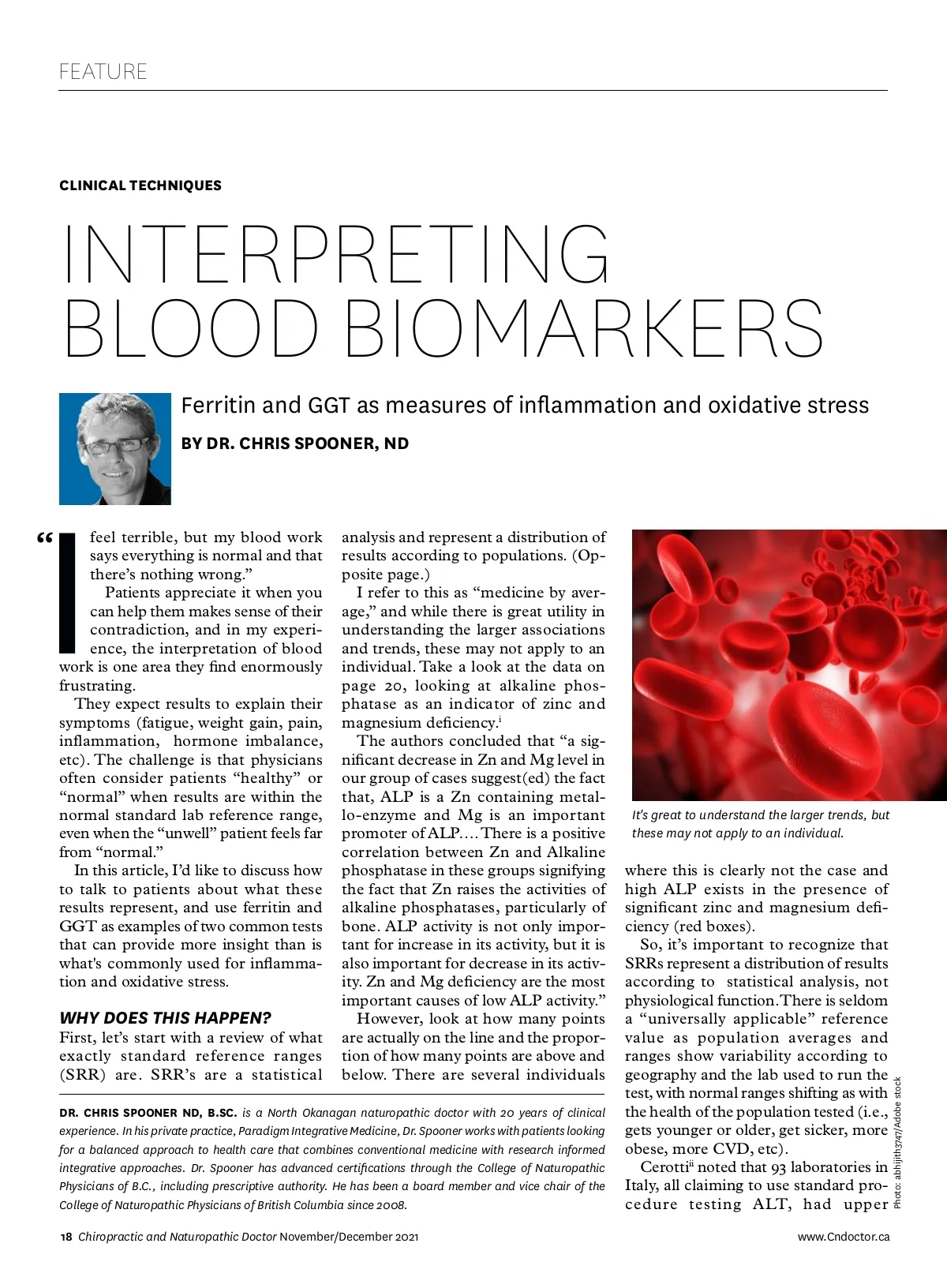
2.5% percentile 2.5% Reference Population 95% 97.5% percentile 2.5% “Not Normal” Percentile 1 5 10 20 “Normal” 30 40 50 60 70 Cententration 80 90 95 99 “Not Normal” A review of what Standard Reference Ranges are. reference limits for adult males span-ning from 40 U/L to 72 U/L, while the lower limit ranged from 0 U/L to 30 U/L. They concluded that: “This com-mon situation is dangerous and mis-leading both for clinicians and patients (the same analytical result can be considered ‘normal’ in one laboratory and ‘abnormal’ in another, according to the reference interval in use).” ALT is an important marker for measuring liver damage and is commonly used to monitor liver damage associated with numerous medications. This leads to a situation where stand-ard reference ranges are basically showing you what’s average and not optimal and where the test may not be sensitive enough to detect disease early and only detect overt pathology once the damage is done. To summarize, the “standard” ap-proach is using laboratory results as guideposts for diagnosing and moni-toring disease where “out of range” results indicate the presence of a dis-ease or condition associated with the biomarker and “within range” results indicate the absence of disease – the absence of disease is synonymous with “health.” Consequently, a more pragmatic approach is needed, and health should be judged subjectively as the absence of signs of disease specifically related to the measurand. Reviewing that same blood work in a context of “optimal ranges” may demonstrate that the pa-tient is far from “normal” by looking at a narrower range of where results are associated with optimal health not just the absence of disease, can detect early changes and trends away from optimal www.Cndoctor.ca function, and provide an opportunity to further explore and assess potential dysfunction before it progresses to disease. This is obviously a very large topic, and many integrative clinicians are currently using the optimal range ap-proach, TSH testing is the most obvious case I can think of. However, I find I am using this approach much more fre-quently with ferritin and GGT so will lay out the case for each of these tests. Serum ferritin levels are widely meas-ured as indicators of iron status. In clinical practice, I see low ferritin ben used to justify iron supplementation. Even in the context of normal RBC counts, hematocrit, and mean corpus-cular volume. When it is elevated, the most interpretation is hemochromato-sis and therapy involves monitoring of liver enzymes and, if significantly ele-vated, phlebotomy. To review, ferric (+3) salts and ions are poorly water-soluble and much of the complex (redox) chemistry of iron in the body is designed to deal with this. iii Iron is absorbed in the intestine as ferrous (+2) ions and transported in the serum bound (in the ferric form, +3) to transferrin, where it can enter peripheral tissues via suitable recep-tors, being re-reduced in the process (See page 21). iv However, “serum ferritin” presents a paradox, as the iron v storage protein ferritin is not synthesized in serum yet is found there. Ferritin is supposed to be a cellular means of storing iron not of transporting it, yet serum ferritin levels are widely measured as indica-FERRITIN tors of iron status. A study of iron deficiency in female athletes vi found that even though serum iron levels of athletes were higher, se-rum ferritin levels were lower than in control subjects and physical activity study with 1743 Finnish men, the du-ration and frequency of physical activ-ity were associated inversely with serum ferritin and blood hemoglobin. vii Ferritin is the necessary protein that binds iron to stop its otherwise excep-tional reactivity, specifically the pro-duction of the very damaging hydroxyl radical that reacts in nanoseconds with the nearest biological substances mak-ing it especially dangerous. viii Thus, while iron is vital for living processes, there is an exceptionally important need to sequester iron in a suitably li-ganded form, and cellular ferritin is a major means of doing this. Serum ferritin is also a well-known inflammatory marker and levels can be raised significantly in response to in-flammation and/or a variety of diseases. It appears that serum ferritin reflects or causes inflammation, and that se-rum ferritin arises from damaged cells, and is thus a marker of cellular damage. The protein in serum ferritin is consid-ered benign, but it has lost (i.e. dumped) most of its normal comple-ment of iron which when unliganded is highly toxic. In this context, it would be prudent to re-evaluate ferritin and iron status in the context of other functional mark-ers. For low ferritin RBC, Hematocrit, Hemoglobin, MCHC and MCV should be considered. ix In cases of ele-vated ferritin, recall that serum ferritin likely originates from cell leakage and that there is a correlation between se-rum aspartate aminotransferase (a marker of hepatocellular damage) and SF. Serum alanine aminotransferase is another well-known marker of liver damage that correlates with serum ferritin, consistent with the view that serum ferritin is indeed a marker of damaged cells. x Looking at ferritin in this context opens up numerous op-tions for therapy such as antioxidant support and hepatic support. GAMMA GLUTAMYL TRANSFERASE (GGT) Gamma-glutamyltransferase (GGT) is a well-established serum marker for November/December 2021 Chiropractic and Naturopathic Doctor 19
Chiropractic + Naturopathic Doctor November/December 2021: Page 19