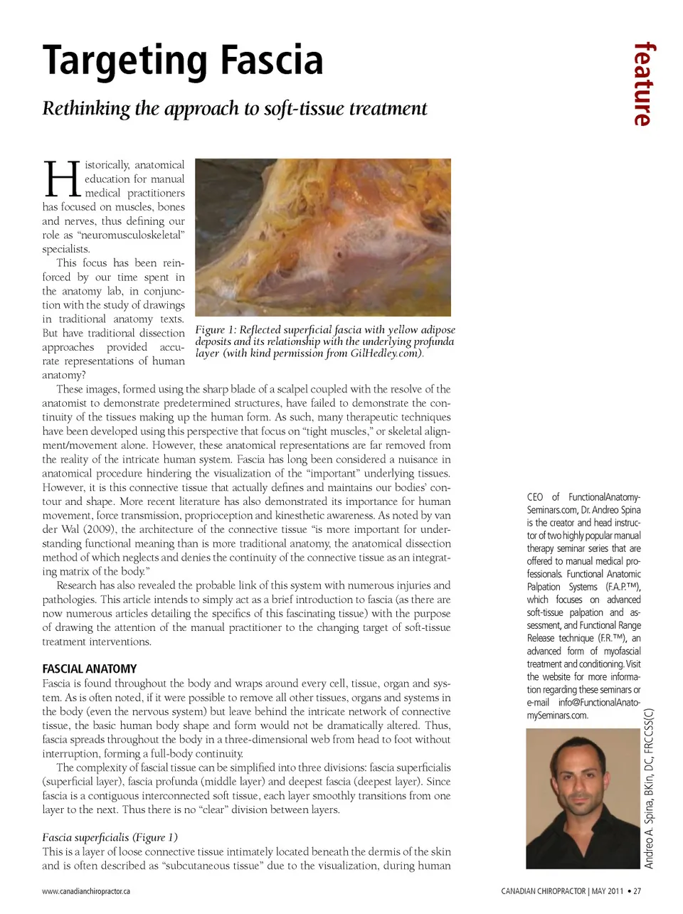
Targeting Fascia Rethinking the approach to soft-tissue treatment feature H istorically, anatomical education for manual medical practitioners has focused on muscles, bones and nerves, thus defining our role as “neuromusculoskeletal” specialists. This focus has been rein-forced by our time spent in the anatomy lab, in conjunc-tion with the study of drawings in traditional anatomy texts. But have traditional dissection Figure 1: Reflected superficial fascia with yellow adipose approaches provided accu-deposits and its relationship with the underlying profunda layer (with kind permission from GilHedley.com). rate representations of human anatomy? These images, formed using the sharp blade of a scalpel coupled with the resolve of the anatomist to demonstrate predetermined structures, have failed to demonstrate the con-tinuity of the tissues making up the human form. As such, many therapeutic techniques have been developed using this perspective that focus on “tight muscles,” or skeletal align-ment/movement alone. However, these anatomical representations are far removed from the reality of the intricate human system. Fascia has long been considered a nuisance in anatomical procedure hindering the visualization of the “important” underlying tissues. However, it is this connective tissue that actually defines and maintains our bodies’ con-tour and shape. More recent literature has also demonstrated its importance for human movement, force transmission, proprioception and kinesthetic awareness. As noted by van der Wal (2009), the architecture of the connective tissue “is more important for under-standing functional meaning than is more traditional anatomy, the anatomical dissection method of which neglects and denies the continuity of the connective tissue as an integrat-ing matrix of the body.” Research has also revealed the probable link of this system with numerous injuries and pathologies. This article intends to simply act as a brief introduction to fascia (as there are now numerous articles detailing the specifics of this fascinating tissue) with the purpose of drawing the attention of the manual practitioner to the changing target of soft-tissue treatment interventions. Fascia superficialis (Figure 1) This is a layer of loose connective tissue intimately located beneath the dermis of the skin and is often described as “subcutaneous tissue” due to the visualization, during human www.canadianchiropractor.ca CANAdiAN CHiROpRACTOR | MAY 2011 • 27 Andreo A. Spina, BKin, dC, FRCCSS(C) FASCIAL ANATOMY Fascia is found throughout the body and wraps around every cell, tissue, organ and sys-tem. As is often noted, if it were possible to remove all other tissues, organs and systems in the body (even the nervous system) but leave behind the intricate network of connective tissue, the basic human body shape and form would not be dramatically altered. Thus, fascia spreads throughout the body in a three-dimensional web from head to foot without interruption, forming a full-body continuity. The complexity of fascial tissue can be simplified into three divisions: fascia superficialis (superficial layer), fascia profunda (middle layer) and deepest fascia (deepest layer). Since fascia is a contiguous interconnected soft tissue, each layer smoothly transitions from one layer to the next. Thus there is no “clear” division between layers. CEO of FunctionalAnatomy-Seminars.com, Dr. Andreo Spina is the creator and head instruc-tor of two highly popular manual therapy seminar series that are offered to manual medical pro-fessionals. Functional Anatomic palpation Systems (F.A.p.™), which focuses on advanced soft-tissue palpation and as-sessment, and Functional Range Release technique (F.R.™), an advanced form of myofascial treatment and conditioning. Visit the website for more informa-tion regarding these seminars or e-mail [email protected].
Chiropractic + Naturopathic Doctor May 2011: Page 27