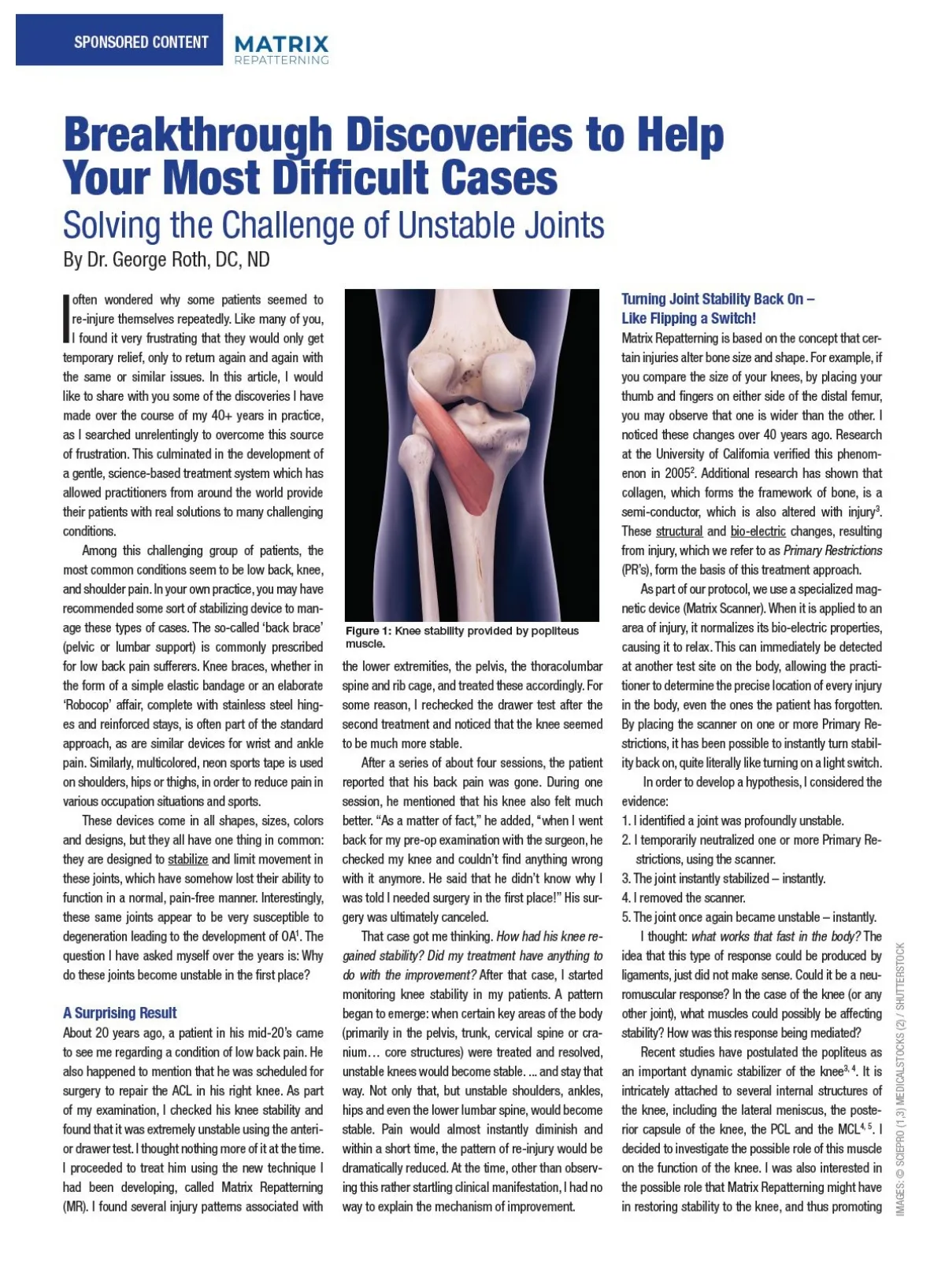
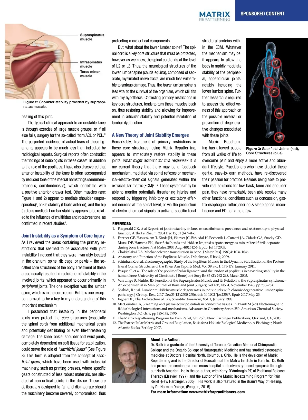
SPONSORED CONTENT SPONSORED CONTENT SPONSORED CONTENTars R L patients, as well as participating in the training program himself. In his recent best-selling protecting more critical components. structural proteins with-book, The Brain’s Way of Healing, he comments: “I view it as prudent to have Matrix assessment Subluxation: A case of mistaken identity? But, a what about the lower lumbar spine? The spi-in the ECM. Whatever after a blow to the head… observing such cases nal cord is a key core structure that must be protected, the mechanism may be, has led me to hope By that Dr. soon, Matrix Repat-George Roth however applied as we know, the spinal cord ends at the level it appears to allow the terning will be routinely in hospital 2 emergency departments.” of L2 or L3. Thus, the neurological structures of the body to rapidly modulate Observations in a new light to e and roved bones oned. e may mean igned ent of ure 1, hieve may cture tently, on, if t that n the would more hieve me to hasis rvous , as a area ional . The ne is l pro-owing was al-r any rding urons ective nately, most of the references emphasized While studying radiology in chiropractic ing and evaluation measurable, objective ble of to the serious damage. Thus, the lower lumbar spine is notably including the lack of scientific validation or measur-school, I noticed that the size of a structure on changes (biomechanical, structural, radio-spine. Fur-less vital to the survival of the which fits side lower able outcomes to support the basic tenets of still one of the lumbar body was often different than graphic, biochemical and neurological) in my organism, our profession. At best, there are a few studies its in counterpart on the other side. For example, A Testable Hypothesis for a Mole ther research is required my Correcting primary restrictions own practice and with those of hypothesis. my students and Figure 4, Post-treatment: Femoral and that demonstrate a modest advantage for chi-the proximal femoral or humeral head, or the Fracture Resistance Figure 2: Shoulder stability provided by supraspi-colleagues, I was key gradually able to evolve core structures, tends a to turn these muscles back to assess the effective-tibial epiphyses restored to same size as ropractic manipulation over some prescription tibial plateau on one side was noticeably larger A natus muscle. method to identify and resolve many of the the right side, following treatment. *Note of this on individual. on, thus effects restoring stability and allowing for as improve-medications for certain such than the ness opposite side approach in the same osseous (and other fascial) of injury and conditions, opening of the medial joint space. back However, impression is My of professors were unable to shed or any light on the possible reversal in pain. articular stability and potential resolution healing of this joint. thus improve our ment clinical outcomes. Over the the overall that been chiropractic science As an anatomy lab instructor at years, this protocol has recognized by a is still relatively un-these findings. prevention of degenera-lumbar dysfunction. The typical clinical approach to an unstable knee and that are only mini-the college and from subsequent observations structure of the cranium. It was only when I growing number of proven chiropractors, as chiropractors well as tive changes associated is through exercise of large medicine muscle groups, or if and all researchers mally from accepted as part of I the health care team. of cadaverous specimens, I was able to confirm B embarked on a study of osteopathic clinicians various fields. I have been a of chiropractor for over 40 years, these differences. At the time, these facts were with these joints. else fails, surgery for this the omission so-called “torn ACL PCL.” A to New Theory Emerges that I realized how profound was. have or been gratified note that many of Joint them Stability but early the in my career, I became convinced that filed away, and it was only many years later that The more investigated this important aspect been lig-able to Remarkably, reproduce same measur-Matrix Repattern-The I purported incidence of actual tears have of these treatment of primary restrictions in I I was not achieving the kinds of results prom-these early observations came to be viewed in a of human anatomy and the common injuries, able outcomes, which observed. ing has allowed people these core structures, using Matrix Repatterning, aments appears to be much less than indicated by Figure 3: Sacrificial Joints (red), ised when I attended school. I also witnessed new light, based on my clinical research. which can often lead to life-altering outcomes, My goal has always been to find measurable Core Structures (blue). many of my colleagues By carefully a better quality skel-from examining all walks of life to appears to immediately stability in these radiological reports. Surgical it reports contradict the more I recognized how important was for often evidence to support any of the techniques we, becoming as restore disheartened C or I failing in practice. They had come into this etal model, which is cast from a real skeleton, 6 me to the incorporate a rational approach to the have wondered joints. findings of radiologists in these cases chiropractors, . In addition provide. What often might account for this response? It is overcome pain and enjoy a more active and abun-profession with high expectations and a sincere you can verify many of these same discrepan-treatment of this area. how things might be different if our profession dant lifestyle. have studied these to the role of the popliteus, I have also discovered that my integrate current theory that there may be feedback to help their fellow humans, but a the cies for yourself. Besides Practitioners the examples who of the By applying the principle of identifying were to embrace and desire the latest devel-types of treatments they were taught did or not femur, tibia and easy-to-learn humerus mentioned earlier, gentle, methods, have re-discovered anterior instability of the is often accompanied mechanism, mediated via spinal reflexes mechan-and normalizing the structure of knee the cranial opments in cell biology, molecular biomechan-live up to these expectations. a close inspection of the spine can be very re-4, bones, by as reduced with other areas the body, we have ics, biomedical engineering and bio-electricity tone of of the medial hamstrings (semimem-ical-electro-chemical signals generated within the their passion for practice. Besides being able to pro-Like many of you, I pursued a long search vealing. Note the differences in the size (width, D 5 . These emerging disciplines witnessed a remarkable degree of success in are crucial to our 9 -12 vide real of solutions for processes) low back, at knee and shoulder branosus, semitendinosus), which correlates with extracellular matrix (ECM) to improve . These my systems be and for additional modalities results, may depth height the articular helping individuals recover from many dev-understanding of the effects of injury at the most my body. outcomes and threatening give me the confi-the spine. Figure pain, throughout they have remarkably been 1 able resolve many a positive anterior drawer test. Other muscles (see: monitor potentially injuries various and levels astating neurological consequences, including fundamental level able of validate the to human It is my dence to be able to find and resolve my patient’s demonstrates these differences in the size of cognitive, visual, auditory, vestibular and neu-belief that these scientific advances would sup-Figure 1 and 2) appear to mediate shoulder (supra-respond by triggering inhibitory or excitatory effer-other functional conditions such as concussion, gas-conditions. In this search, romuscular conditions. Several independent port much of what we already provide and help I was blessed to meet the articular processes of the atlas. I contend Figure 2: Microscopic Evidence tro-esophageal reflux, & sleep apnea, incon-spinatus) 7 , ankle stability (tibialis anterior), neurons at the spinal level, or via the production and the hip ent several researchers and clinicians from other that on palpation, these areas of snoring enlargement researchers have verified these outcomes. 3, 4 us evolve even further as a truly science-based Expansion with Injury, Paul Han fields (cell biology, biomedical engineering, may be easily mistaken for a relative rotation or of electro-chemical signals to activate specific tonal tinence and ED, to name a few. Lumbar appears to be relat-profession. I sense that there is a growing Dr. (gluteus Norman medius). Doidge, MD, who stability is on faculty University of California, Berkele medicine, and physical translation of the vertebral segment (see: Sub-permission from the author) desire tone, among in our profession to osteopathy see at Columbia University and the University ed to the influence of multifidus and of rotatores as many orthopedic medicine). were making amazing discov-luxation: A Case of Mistaken Identity, below). role They in the health Toronto, is a world-renowned expert in the chiropractic take its rightful REFERENCES: confirmed in recent studies 8 . eries the underlying effects of injury Recent evidence from the University of Cal-male hockey player, who had in regarding the field of physical field of brain injury and neuroplasticity. After care industry, as leaders 1. and Fitzgerald GK, et al Reports of at joint knee osteoarthritis: its powerful prevalence and relationship to physical biomechanical dysfunction the instability cellular, in ifornia, revealed by the Atomic Force from knee pain for several m medicine. The way ahead benefits not only our hearing about our approach, he conducted an function, Arthritis Rheum. 2004 Dec 15; 51 (6): bio-electrical even the molecular level. I 941-6. microscope, under the direction of physicist prevented him from playing. T profession but also the countless and individuals in-depth investigation, which included obser-Joint Instability as a Symptom of Core Injury 2. Fantner GE, Hassenkam T, Kindt JH, Weaver JC, Birkedal H, and Pechenik L, Cutroni JA, Cidade Stucky GD, 1 , has confirmed that for many a system of therapeutics Paul Hansma his team caused considerable pain durin the GA, real solutions for vations of treatments, patient interviews with who are looking for recognized Morse DE, Hansma Sacrificial with bonds and hidden length dissipate energy as mineralized separate As I reviewed the areas containing the primary re-to be valid, it had to PK be , congruent this activities, such as climbing sta presence of certain protein structures within fibrils numerous concussion and post-concussion painful and limiting conditions. during bone fracture, Nat Mater. 2005 Aug; 4(8):612-6. Epub. Jul 17 2005. emerging science. the bone that expand with an injury. These was only able to accomplish i strictions that seemed to be associated with joint 3. Chakkalakal DA, Mechanoelectric transduction in bone. are J Mater Res]. 1989;4: 1034-1046. consistent with my clinical obser-bling manner. Fortunately for m instability, I noticed that they were invariably located 4. Anatomy and Function of the Popliteus Muscle, findings I MacIntyre, E-book, 2009. References: surgeons were monitoring the si vations, which were first made over 40 years Popliteus Muscle of the the Postero-in the cranium, spine, rib cage, or pelvis – the so-5. Schinhan K, et al, Electromyographic Study of the knee with a high degre ago (see Figure 2). in the Dynamic Stabilization at 1. Fantner GE, Hassenkam T, Kindt JH, Weaver JC, Birkedal H, Pechenik L, Cutroni JA, Corner Structures of the Knee, Am J Sports Med, Vol. 39, no. 1, 173-179, January, 2011. Cidade GA, Stucky GD, of Morse DE, Hansma PK, Sacrificial and hidden length due to an underlying genetic c called core structures the body. Treatment of these bonds lateral dissipate energy as mineralized fibrils separate during bone 6. fracture, Nat Pasque C, Mater. et al, The role of the popliteofibular ligament and the tendon of popliteus in providing stability in the result, they took consistent me Restoration of Bone Size 2005 Aug;4(8):612-6. Epub Jul 17. of stability in the areas usually resulted in 2005 restoration human knee, University of Cincinnati, J Bone Joint Surg Br. 85 (2): 292-298, March 2003. within one-hundredth of a m and Joint Healing 2. involved Doidge, N., The Brain’s of Healing, Penguin Books, York, 2016. joints, which Way appeared to occur primarily in New 7. Van Linge B, Mulder JD, Function of the Supraspinatus Muscle and its clinical Relation to the Supraspinatus -when the fe were syndrome all surprised One of the significant breakthroughs experimental Vol 45B, and No. 4, pp 750–754. 3. Tommerdahl, M, Dennis, RG, et al., Neurosensory Assessment of An Concussion, Mil in Man, Journal of Bone and Joint and the tibial plateau of the le that Surgery, my colleagues I November were able 1963, to ac-peripheral joints. The one exception was the lumbar Med. 2016 May;181(5 Suppl):45-50 8. Shahidi, B et al, Lumbar multifidus muscle degenerates individuals with chronic degenerative lumbar been spine approximately 5 mm complish in was that bone size appeared to be had which LA, is in the core and region. But this one excep-in connective 4. spine, MacGuintie Streaming piezoelectric potentials tissues, In: pathology, J Orthop. Res., 2017 Dec;35(12):2700-2706. doi: 10.1002/jor.23597. Epub 2017 May 23. counterpart on the right, had su restored to normal with treatment. At first, we Blank M (ed) Electromagnetic fields: biological interactions and mechanisms. Vol. 1, January 1998. tion, proved to be a key to my understanding of this 9. Ingber DE, The Architecture of Life, Scientific American, by that amount after only a few questioned these results and followed them up Advances in Chemistry Series 250. American Chemical Society, Washington DC, ch. 10. MacGuintie LA, Streaming and piezoelectric potentials in connective tissues, In: Blank M and (ed) Electromagnetic Figure 3 and 4). Subsequently, th with precise measurements using callipers 8, pp 125-142, 1995. important mechanism. Figure 1: Model of C1, cast from a real fields: biological interactions and mechanisms. Advances in Chemistry Series 250. American Society, parents noted that he was onc tape measures. Inter-tester validation appeared Chemical specimen. Note the enlargement 5. Chakkalakal DA, Mechanoelectric transduction in bone. J Mater skeletal Res]. 1989;4: 1034-1046. I postulated that instability in the peripheral Washington DC, ch. 8, pp 125-142, 1995. of the right articular process and the spinal up” the stairs with absolutely no to confirm our findings. canal, which measured 2 mm. larger in joints may protect the For 11. The Matrix Repatterning Program for Pain Relief, GB Roth, New ago, Harbinger Publications, Oakland, CA, 2005. core structures (especially verified my contention that no Several years I treated a 15-year-old more information: matrixforpractitioners.com every dimension. the spinal cord) from additional mechanical strain 12. The Extracellular Matrix and Ground Regulation, Basis for a Holistic Biological Medicine, A Pischinger, North Atlantic Books, Berkley, 2007. lower lumbar searched spine (cauda equina), composed stability of the peripher-recently the Internet for scientific of sep-Startling Observations: The Evolution of Chiropractic Bone Enlarges with Injury! to support chiropractic. Unfortu-arate, evidence myelinated nerve tracts, are much less vulnera-al, appendicular joints, Through a combination of relentless question-I and potentially debilitating or even life-threatening damage. The knee, ankle, shoulder and wrist joints, About the Author: completely dependent on soft tissue for stabilization, Dr. Roth is a graduate of the University of Toronto, Canadian Memorial Chiropractic CC_Matrix_Sponsored_Content_DPS_Dec19_EJS.indd CC_Dec19_EJS.indd 18 2019-11-06 8:38 AM 2-3 could serve the role of “sacrificial joints” (See Figure College and the Ontario College of Naturopathic Medicine and has studied osteopathic medicine at Doctors’ Hospital North, Columbus, Ohio. He is the developer of Matrix 3). This term is adapted from the concept of sacri-Repatterning and is the Director of Education at the Matrix Institute in Toronto. Dr. Roth ficial gears, which have been used with industrial has presented seminars at numerous hospital and university-based symposia through-machinery, such as printing presses, where specific out North America. He is the co-author, with Kerry D’Ambrogio PT, of Positional Release gears constructed of less robust materials, are situ-Therapy (Elsevier, 1997), and the author of The Matrix Repatterning Program for Pain ated at non-critical points in the device. These are Relief (New Harbinger, 2005). His work is also featured in the Brain’s Way of Healing, by Dr. Norman Doidge, (Penguin, 2015). deliberately designed to fail and disintegrate should For more information: wwwmatrixforpractitioners.com the machinery become severely compromised, thus
Chiropractic + Naturopathic Doctor December 2020: Page 19