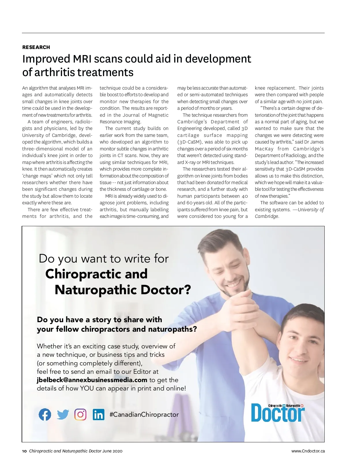
RESEARCH Improved MRI scans could aid in development of arthritis treatments An algorithm that analyses MRI im-ages and automatically detects small changes in knee joints over time could be used in the develop-ment of new treatments for arthritis. A team of engineers, radiolo-gists and physicians, led by the University of Cambridge, devel-oped the algorithm, which builds a three-dimensional model of an individual’s knee joint in order to map where arthritis is affecting the knee. It then automatically creates ‘change maps’ which not only tell researchers whether there have been significant changes during the study but allow them to locate exactly where these are. There are few effective treat-ments for arthritis, and the technique could be a considera-ble boost to efforts to develop and monitor new therapies for the condition. The results are report-ed in the Journal of Magnetic Resonance Imaging. The current study builds on earlier work from the same team, who developed an algorithm to monitor subtle changes in arthritic joints in CT scans. Now, they are using similar techniques for MRI, which provides more complete in-formation about the composition of tissue --not just information about the thickness of cartilage or bone. MRI is already widely used to di-agnose joint problems, including arthritis, but manually labelling each image is time-consuming, and may be less accurate than automat-ed or semi-automated techniques when detecting small changes over a period of months or years. The technique researchers from Cambridge’s Department of Engineering developed, called 3D cartilage surface mapping (3D-CaSM), was able to pick up changes over a period of six months that weren’t detected using stand-ard X-ray or MRI techniques. The researchers tested their al-gorithm on knee joints from bodies that had been donated for medical research, and a further study with human participants between 40 and 60 years old. All of the partic-ipants suffered from knee pain, but were considered too young for a knee replacement. Their joints were then compared with people of a similar age with no joint pain. “There’s a certain degree of de-terioration of the joint that happens as a normal part of aging, but we wanted to make sure that the changes we were detecting were caused by arthritis,” said Dr James MacKay from Cambridge’s Department of Radiology, and the study’s lead author. “The increased sensitivity that 3D-CaSM provides allows us to make this distinction, which we hope will make it a valua-ble tool for testing the effectiveness of new therapies.” The software can be added to existing systems. —University of Cambridge. Do you want to write for Chiropractic and Naturopathic Doctor? Do you have a story to share with your fellow chiropractors and naturopaths? Whether it’s an exciting case study, overview of a new technique, or business tips and tricks (or something completely different), feel free to send an email to our Editor at [email protected] to get the details of how YOU can appear in print and online! #CanadianChiropractor 10 Chiropractic and Naturopathic Doctor June 2020 CC_HouseAdwritefor_May20_MLD.indd 1 www.Cndoctor.ca 2020-04-14 10:18 AM
Chiropractic + Naturopathic Doctor June 2020: Page 10