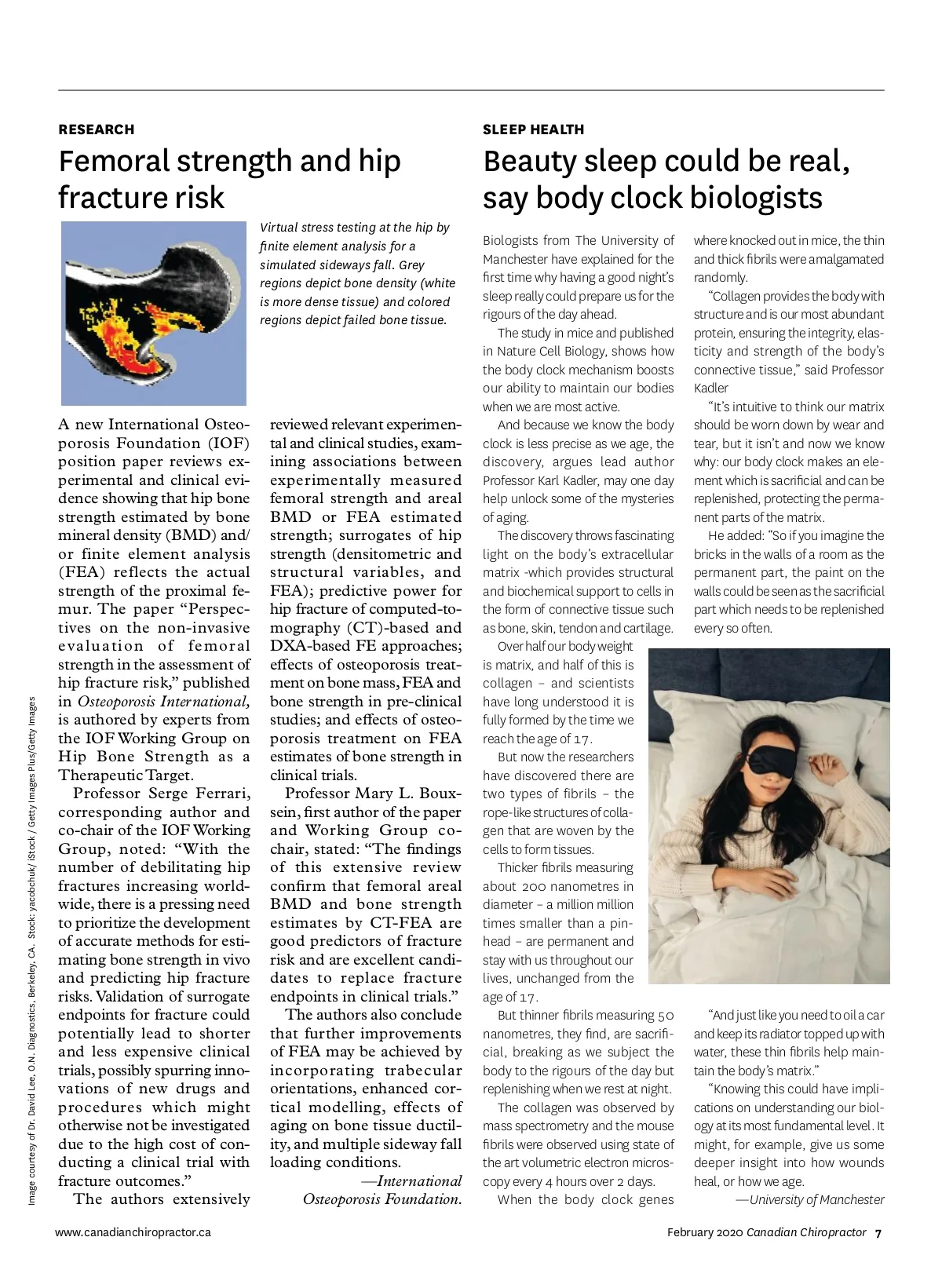
RESEARCH Femoral strength and hip fracture risk Virtual stress testing at the hip by finite element analysis for a simulated sideways fall. Grey regions depict bone density (white is more dense tissue) and colored regions depict failed bone tissue. SLEEP HEALTH Beauty sleep could be real, say body clock biologists Biologists from The University of Manchester have explained for the first time why having a good night’s sleep really could prepare us for the rigours of the day ahead. The study in mice and published in Nature Cell Biology, shows how the body clock mechanism boosts our ability to maintain our bodies when we are most active. And because we know the body clock is less precise as we age, the discovery, argues lead author Professor Karl Kadler, may one day help unlock some of the mysteries of aging. The discovery throws fascinating light on the body’s extracellular matrix -which provides structural and biochemical support to cells in the form of connective tissue such as bone, skin, tendon and cartilage. Over half our body weight is matrix, and half of this is collagen – and scientists have long understood it is fully formed by the time we reach the age of 17. But now the researchers have discovered there are two types of fibrils – the rope-like structures of colla-gen that are woven by the cells to form tissues. Thicker fibrils measuring about 200 nanometres in diameter – a million million times smaller than a pin-head – are permanent and stay with us throughout our lives, unchanged from the age of 17. But thinner fibrils measuring 50 nanometres, they find, are sacrifi-cial, breaking as we subject the body to the rigours of the day but replenishing when we rest at night. The collagen was observed by mass spectrometry and the mouse fibrils were observed using state of the art volumetric electron micros-copy every 4 hours over 2 days. When the body clock genes where knocked out in mice, the thin and thick fibrils were amalgamated randomly. “Collagen provides the body with structure and is our most abundant protein, ensuring the integrity, elas-ticity and strength of the body’s connective tissue,” said Professor Kadler “It’s intuitive to think our matrix should be worn down by wear and tear, but it isn’t and now we know why: our body clock makes an ele-ment which is sacrificial and can be replenished, protecting the perma-nent parts of the matrix. He added: “So if you imagine the bricks in the walls of a room as the permanent part, the paint on the walls could be seen as the sacrificial part which needs to be replenished every so often. A new International Osteo-porosis Foundation (IOF) position paper reviews ex-perimental and clinical evi-dence showing that hip bone strength estimated by bone mineral density (BMD) and/ or finite element analysis (FEA) reflects the actual strength of the proximal fe-mur. The paper “Perspec-tives on the non-invasive evaluation of femoral strength in the assessment of hip fracture risk,” published in Osteoporosis International, is authored by experts from the IOF Working Group on Hip Bone Strength as a Therapeutic Target. Professor Serge Ferrari, corresponding author and co-chair of the IOF Working Group, noted: “With the number of debilitating hip fractures increasing world-wide, there is a pressing need to prioritize the development of accurate methods for esti-mating bone strength in vivo and predicting hip fracture risks. Validation of surrogate endpoints for fracture could potentially lead to shorter and less expensive clinical trials, possibly spurring inno-vations of new drugs and procedures which might otherwise not be investigated due to the high cost of con-ducting a clinical trial with fracture outcomes.” The authors extensively www.canadianchiropractor.ca reviewed relevant experimen-tal and clinical studies, exam-ining associations between experimentally measured femoral strength and areal BMD or FEA estimated strength; surrogates of hip strength (densitometric and structural variables, and FEA); predictive power for hip fracture of computed-to-mography (CT)-based and DXA-based FE approaches; effects of osteoporosis treat-ment on bone mass, FEA and bone strength in pre-clinical studies; and effects of osteo-porosis treatment on FEA estimates of bone strength in clinical trials. Professor Mary L. Boux-sein, first author of the paper and Working Group co-chair, stated: “The findings of this extensive review confirm that femoral areal BMD and bone strength estimates by CT-FEA are good predictors of fracture risk and are excellent candi-dates to replace fracture endpoints in clinical trials.” The authors also conclude that further improvements of FEA may be achieved by incorporating trabecular orientations, enhanced cor-tical modelling, effects of aging on bone tissue ductil-ity, and multiple sideway fall loading conditions. —International Osteoporosis Foundation. Image courtesy of Dr. David Lee, O.N. Diagnostics, Berkeley, CA. Stock: yacobchuk/ iStock / Getty Images Plus/Getty Images “And just like you need to oil a car and keep its radiator topped up with water, these thin fibrils help main-tain the body’s matrix.” “Knowing this could have impli-cations on understanding our biol-ogy at its most fundamental level. It might, for example, give us some deeper insight into how wounds heal, or how we age. —University of Manchester February 2020 Canadian Chiropractor 7
Chiropractic + Naturopathic Doctor February 2020: Page 7