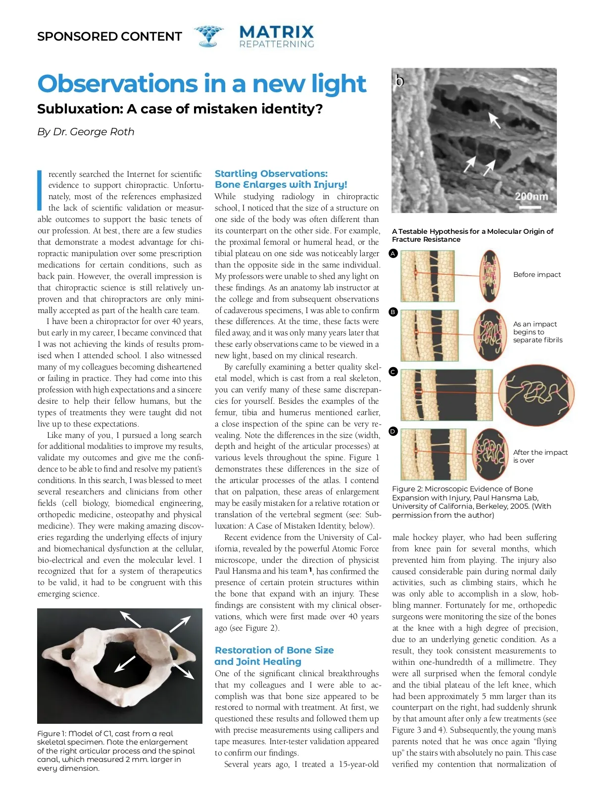
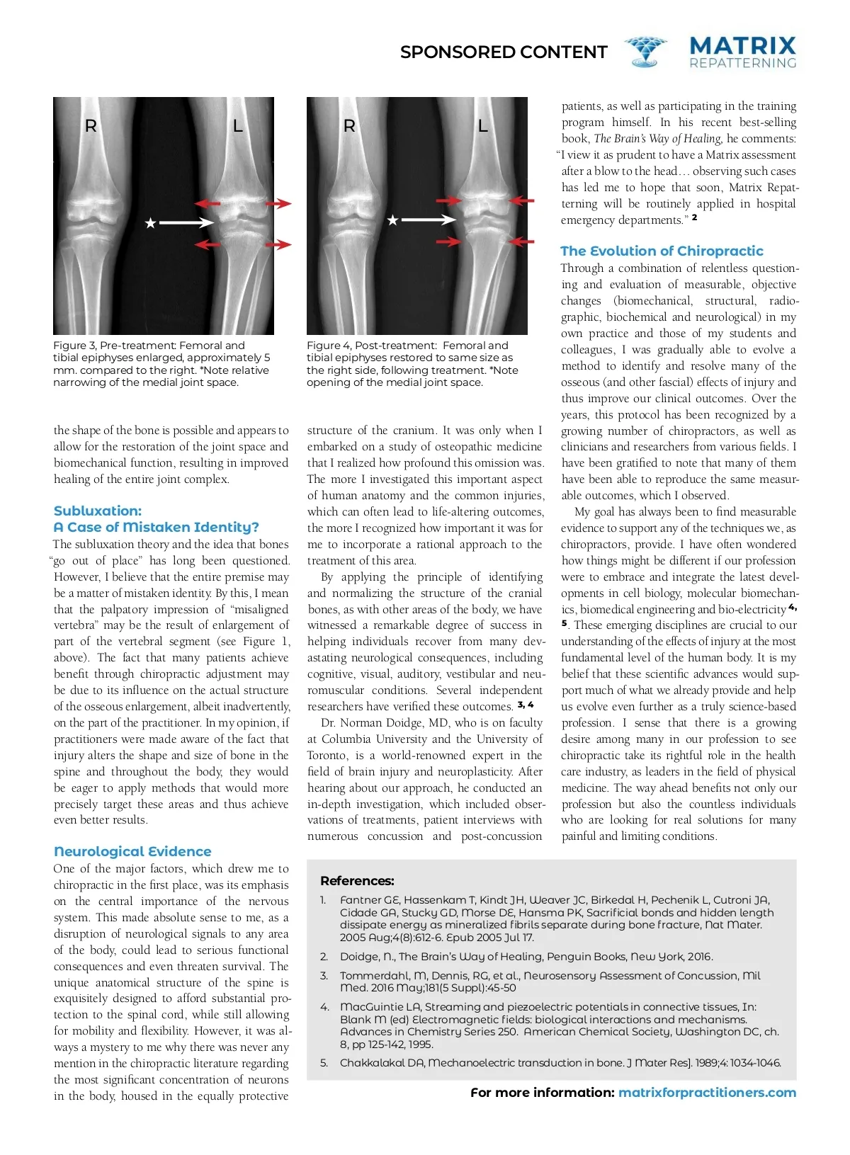
SPONSORED CONTENT patients, as well as participating in the training program himself. In his recent best-selling book, The Brain’s Way of Healing, he comments: “I view it as prudent to have a Matrix assessment after a blow to the head… observing such cases has led me to hope that soon, Matrix Repat-terning will be routinely applied in hospital emergency departments.” 2 R L R L The Evolution of Chiropractic Through a combination of relentless question-ing and evaluation of measurable, objective changes (biomechanical, structural, radio-graphic, biochemical and neurological) in my own practice and those of my students and colleagues, I was gradually able to evolve a method to identify and resolve many of the osseous (and other fascial) effects of injury and thus improve our clinical outcomes. Over the years, this protocol has been recognized by a growing number of chiropractors, as well as clinicians and researchers from various fields. I have been gratified to note that many of them have been able to reproduce the same measur-able outcomes, which I observed. My goal has always been to find measurable evidence to support any of the techniques we, as chiropractors, provide. I have often wondered how things might be different if our profession were to embrace and integrate the latest devel-opments in cell biology, molecular biomechan-ics, biomedical engineering and bio-electricity 4, 5 . These emerging disciplines are crucial to our understanding of the effects of injury at the most fundamental level of the human body. It is my belief that these scientific advances would sup-port much of what we already provide and help us evolve even further as a truly science-based profession. I sense that there is a growing desire among many in our profession to see chiropractic take its rightful role in the health care industry, as leaders in the field of physical medicine. The way ahead benefits not only our profession but also the countless individuals who are looking for real solutions for many painful and limiting conditions. Figure 3, Pre-treatment: Femoral and tibial epiphyses enlarged, approximately 5 mm. compared to the right. *Note relative narrowing of the medial joint space. Figure 4, Post-treatment: Femoral and tibial epiphyses restored to same size as the right side, following treatment. *Note opening of the medial joint space. the shape of the bone is possible and appears to allow for the restoration of the joint space and biomechanical function, resulting in improved healing of the entire joint complex. Subluxation: A Case of Mistaken Identity? The subluxation theory and the idea that bones “go out of place” has long been questioned. However, I believe that the entire premise may be a matter of mistaken identity. By this, I mean that the palpatory impression of “misaligned vertebra” may be the result of enlargement of part of the vertebral segment (see Figure 1, above). The fact that many patients achieve benefit through chiropractic adjustment may be due to its influence on the actual structure of the osseous enlargement, albeit inadvertently, on the part of the practitioner. In my opinion, if practitioners were made aware of the fact that injury alters the shape and size of bone in the spine and throughout the body, they would be eager to apply methods that would more precisely target these areas and thus achieve even better results. Neurological Evidence One of the major factors, which drew me to chiropractic in the first place, was its emphasis on the central importance of the nervous system. This made absolute sense to me, as a disruption of neurological signals to any area of the body, could lead to serious functional consequences and even threaten survival. The unique anatomical structure of the spine is exquisitely designed to afford substantial pro-tection to the spinal cord, while still allowing for mobility and flexibility. However, it was al-ways a mystery to me why there was never any mention in the chiropractic literature regarding the most significant concentration of neurons in the body, housed in the equally protective structure of the cranium. It was only when I embarked on a study of osteopathic medicine that I realized how profound this omission was. The more I investigated this important aspect of human anatomy and the common injuries, which can often lead to life-altering outcomes, the more I recognized how important it was for me to incorporate a rational approach to the treatment of this area. By applying the principle of identifying and normalizing the structure of the cranial bones, as with other areas of the body, we have witnessed a remarkable degree of success in helping individuals recover from many dev-astating neurological consequences, including cognitive, visual, auditory, vestibular and neu-romuscular conditions. Several independent researchers have verified these outcomes. 3, 4 Dr. Norman Doidge, MD, who is on faculty at Columbia University and the University of Toronto, is a world-renowned expert in the field of brain injury and neuroplasticity. After hearing about our approach, he conducted an in-depth investigation, which included obser-vations of treatments, patient interviews with numerous concussion and post-concussion References: 1. Fantner GE, Hassenkam T, Kindt JH, Weaver JC, Birkedal H, Pechenik L, Cutroni JA, Cidade GA, Stucky GD, Morse DE, Hansma PK, Sacrificial bonds and hidden length dissipate energy as mineralized fibrils separate during bone fracture, Nat Mater. 2005 Aug;4(8):612-6. Epub 2005 Jul 17. Doidge, N., The Brain’s Way of Healing, Penguin Books, New York, 2016. Tommerdahl, M, Dennis, RG, et al., Neurosensory Assessment of Concussion, Mil Med. 2016 May;181(5 Suppl):45-50 MacGuintie LA, Streaming and piezoelectric potentials in connective tissues, In: Blank M (ed) Electromagnetic fields: biological interactions and mechanisms. Advances in Chemistry Series 250. American Chemical Society, Washington DC, ch. 8, pp 125-142, 1995. Chakkalakal DA, Mechanoelectric transduction in bone. J Mater Res]. 1989;4: 1034-1046. 2. 3. 4. 5. For more information: matrixforpractitioners.com
Chiropractic + Naturopathic Doctor December 2019: Page 19