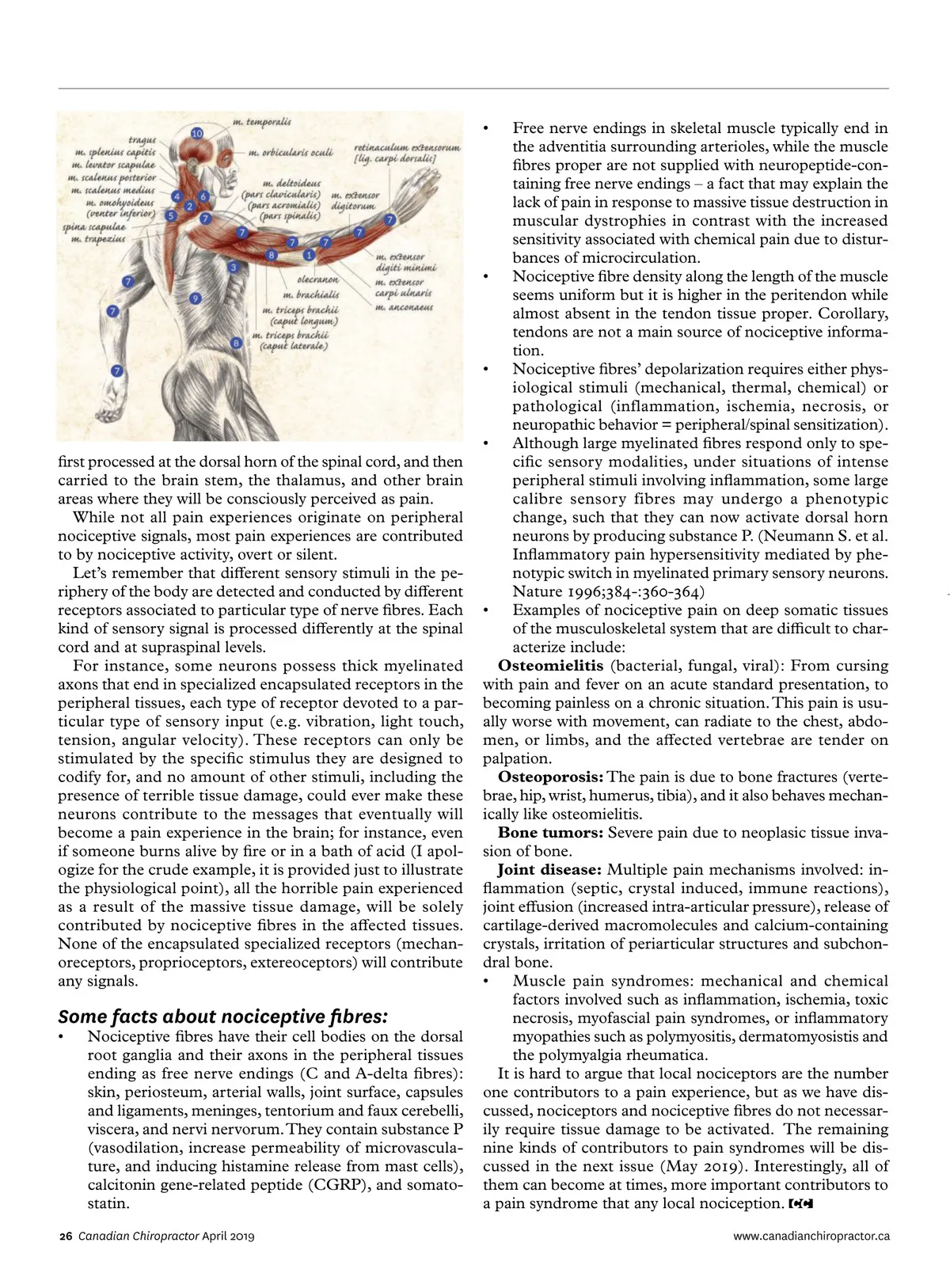
first processed at the dorsal horn of the spinal cord, and then carried to the brain stem, the thalamus, and other brain areas where they will be consciously perceived as pain. While not all pain experiences originate on peripheral nociceptive signals, most pain experiences are contributed to by nociceptive activity, overt or silent. Let’s remember that different sensory stimuli in the pe-riphery of the body are detected and conducted by different receptors associated to particular type of nerve fibres. Each kind of sensory signal is processed differently at the spinal cord and at supraspinal levels. For instance, some neurons possess thick myelinated axons that end in specialized encapsulated receptors in the peripheral tissues, each type of receptor devoted to a par-ticular type of sensory input (e.g. vibration, light touch, tension, angular velocity). These receptors can only be stimulated by the specific stimulus they are designed to codify for, and no amount of other stimuli, including the presence of terrible tissue damage, could ever make these neurons contribute to the messages that eventually will become a pain experience in the brain; for instance, even if someone burns alive by fire or in a bath of acid (I apol-ogize for the crude example, it is provided just to illustrate the physiological point), all the horrible pain experienced as a result of the massive tissue damage, will be solely contributed by nociceptive fibres in the affected tissues. None of the encapsulated specialized receptors (mechan-oreceptors, proprioceptors, extereoceptors) will contribute any signals. Some facts about nociceptive fibres: • Nociceptive fibres have their cell bodies on the dorsal root ganglia and their axons in the peripheral tissues ending as free nerve endings (C and A-delta fibres): skin, periosteum, arterial walls, joint surface, capsules and ligaments, meninges, tentorium and faux cerebelli, viscera, and nervi nervorum. They contain substance P (vasodilation, increase permeability of microvascula-ture, and inducing histamine release from mast cells), calcitonin gene-related peptide (CGRP), and somato-statin. Free nerve endings in skeletal muscle typically end in the adventitia surrounding arterioles, while the muscle fibres proper are not supplied with neuropeptide-con-taining free nerve endings – a fact that may explain the lack of pain in response to massive tissue destruction in muscular dystrophies in contrast with the increased sensitivity associated with chemical pain due to distur-bances of microcirculation. • Nociceptive fibre density along the length of the muscle seems uniform but it is higher in the peritendon while almost absent in the tendon tissue proper. Corollary, tendons are not a main source of nociceptive informa-tion. Nociceptive fibres’ depolarization requires either phys-• iological stimuli (mechanical, thermal, chemical) or pathological (inflammation, ischemia, necrosis, or neuropathic behavior = peripheral/spinal sensitization). • Although large myelinated fibres respond only to spe-cific sensory modalities, under situations of intense peripheral stimuli involving inflammation, some large calibre sensory fibres may undergo a phenotypic change, such that they can now activate dorsal horn neurons by producing substance P. (Neumann S. et al. Inflammatory pain hypersensitivity mediated by phe-notypic switch in myelinated primary sensory neurons. Nature 1996;384-:360-364) Examples of nociceptive pain on deep somatic tissues • of the musculoskeletal system that are difficult to char-acterize include: Osteomielitis (bacterial, fungal, viral): From cursing with pain and fever on an acute standard presentation, to becoming painless on a chronic situation. This pain is usu-ally worse with movement, can radiate to the chest, abdo-men, or limbs, and the affected vertebrae are tender on palpation. Osteoporosis: The pain is due to bone fractures (verte-brae, hip, wrist, humerus, tibia), and it also behaves mechan-ically like osteomielitis. Bone tumors: Severe pain due to neoplasic tissue inva-sion of bone. Joint disease: Multiple pain mechanisms involved: in-flammation (septic, crystal induced, immune reactions), joint effusion (increased intra-articular pressure), release of cartilage-derived macromolecules and calcium-containing crystals, irritation of periarticular structures and subchon-dral bone. • Muscle pain syndromes: mechanical and chemical factors involved such as inflammation, ischemia, toxic necrosis, myofascial pain syndromes, or inflammatory myopathies such as polymyositis, dermatomyosistis and the polymyalgia rheumatica. It is hard to argue that local nociceptors are the number one contributors to a pain experience, but as we have dis-cussed, nociceptors and nociceptive fibres do not necessar-ily require tissue damage to be activated. The remaining nine kinds of contributors to pain syndromes will be dis-cussed in the next issue (May 2019). Interestingly, all of them can become at times, more important contributors to a pain syndrome that any local nociception. • www.canadianchiropractor.ca 26 Canadian Chiropractor April 2019
Chiropractic + Naturopathic Doctor April 2019: Page 26