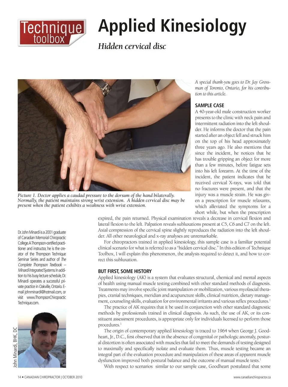
Applied Kinesiology Hidden cervical disc A special thank-you goes to Dr. Jay Gross-man of Toronto, Ontario, for his contribu-tion to this article. Picture 1. Doctor applies a caudad pressure to the dorsum of the hand bilaterally. Normally, the patient maintains strong wrist extension. A hidden cervical disc may be present when the patient exhibits a weakness with wrist extension. SAMPLE CASE A 40-year-old male construction worker presents to the clinic with neck pain and intermittent radiation into the left shoul-der. He informs the doctor that the pain started after an object fell and struck him on the top of his head approximately three years ago. He also mentions that since the incident, he notices that he has trouble gripping an object for more than a few minutes, before fatigue sets into his left forearm. At the time of the incident, the patient indicates that he received cervical X-rays, was told that no fractures were present, and that the injury was a muscle strain. He was giv-en a prescription for muscle relaxants, which alleviated the symptoms for a short while, but when the prescription Dr.John Minardi is a 2001 graduate of Canadian Memorial Chiropractic College.A Thompson-certified practi-tioner and instructor, he is the cre-ator of the Thompson Technique Seminar Series and author of The Complete Thompson Textbook – Minardi Integrated Systems. In addi-tion to his busy lecture schedule,Dr. Minardi operates a successful pri-vate practice in Oakville, Ontario. E-mail [email protected], or visit www.ThompsonChiropractic Technique.com. expired, the pain returned. Physical examination reveals a decrease in cervical flexion and lateral flexion to the left. Palpation reveals subluxations present at C5, C6 and C7 on the left. Axial compression of the cervical spine slightly reproduces the radiation into the left shoul-der. All other neurological and x-ray analyses are unremarkable. For chiropractors trained in applied kinesiology, this sample case is a familiar potential clinical scenario for what is referred to as a “hidden cervical disc.” In this edition of Technique Toolbox, I will explain this phenomenon, the analysis required to detect it, and how to cor-rect this subluxation. BUT FIRST, SOME HISTORY Applied kinesiology (AK) is a system that evaluates structural, chemical and mental aspects of health using manual muscle testing combined with other standard methods of diagnosis. Treatments may involve specific joint manipulation or mobilization, various myofascial thera-pies, cranial techniques, meridian and acupuncture skills, clinical nutrition, dietary manage-ment, counseling skills, evaluation for environmental irritants and various reflex procedures.1 The practice of AK requires that it be used in conjunction with other standard diagnostic methods by professionals trained in clinical diagnosis. As such, the use of AK, or its con-stituent assessment procedures, is appropriate only for individuals licensed to perform those procedures.1 The origin of contemporary applied kinesiology is traced to 1964 when George J. Good-heart, Jr., D.C., first observed that in the absence of congenital or pathologic anomaly, postur-al distortion is often associated with muscles that fail to meet the demands of testing designed to maximally and specifically isolate and evaluate them. Thus, muscle testing became an integral part of the evaluation procedure and manipulation of these areas of apparent muscle dysfunction improved both postural balance and the outcome of manual muscle tests.1 With respect to scenarios similar to our sample case, Goodheart postulated that some 14 • CANADIAN CHIROPRACTOR | OCTOBER 2010 www.canadianchiropractor.ca John Minardi, BHK, DC
Chiropractic + Naturopathic Doctor October 2010: Page 14