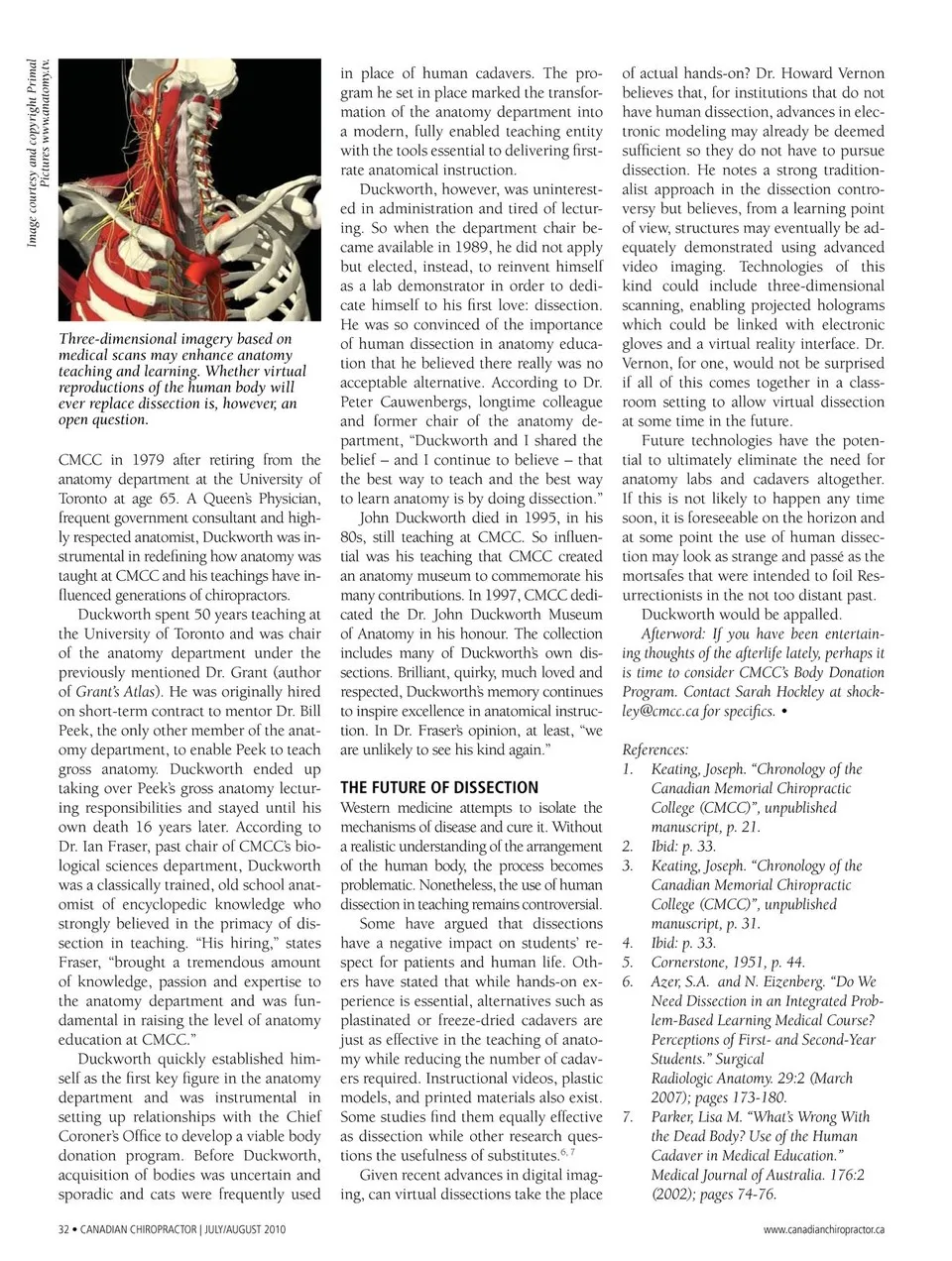
Three-dimensional imagery based on medical scans may enhance anatomy teaching and learning. Whether virtual reproductions of the human body will ever replace dissection is, however, an open question. CMCC in 1979 after retiring from the anatomy department at the University of Toronto at age 65. A Queen’s Physician, frequent government consultant and high-ly respected anatomist, Duckworth was in-strumental in redefining how anatomy was taught at CMCC and his teachings have in-fluenced generations of chiropractors. Duckworth spent 50 years teaching at the University of Toronto and was chair of the anatomy department under the previously mentioned Dr. Grant (author of Grant’s Atlas). He was originally hired on short-term contract to mentor Dr. Bill Peek, the only other member of the anat-omy department, to enable Peek to teach gross anatomy. Duckworth ended up taking over Peek’s gross anatomy lectur-ing responsibilities and stayed until his own death 16 years later. According to Dr. Ian Fraser, past chair of CMCC’s bio-logical sciences department, Duckworth was a classically trained, old school anat-omist of encyclopedic knowledge who strongly believed in the primacy of dis-section in teaching. “His hiring,” states Fraser, “brought a tremendous amount of knowledge, passion and expertise to the anatomy department and was fun-damental in raising the level of anatomy education at CMCC.” Duckworth quickly established him-self as the first key figure in the anatomy department and was instrumental in setting up relationships with the Chief Coroner’s Office to develop a viable body donation program. Before Duckworth, acquisition of bodies was uncertain and sporadic and cats were frequently used 32 • CANADIAN CHIROPRACTOR | JULY/AUGUST 2010 in place of human cadavers. The pro-gram he set in place marked the transfor-mation of the anatomy department into a modern, fully enabled teaching entity with the tools essential to delivering first-rate anatomical instruction. Duckworth, however, was uninterest-ed in administration and tired of lectur-ing. So when the department chair be-came available in 1989, he did not apply but elected, instead, to reinvent himself as a lab demonstrator in order to dedi-cate himself to his first love: dissection. He was so convinced of the importance of human dissection in anatomy educa-tion that he believed there really was no acceptable alternative. According to Dr. Peter Cauwenbergs, longtime colleague and former chair of the anatomy de-partment, “Duckworth and I shared the belief – and I continue to believe – that the best way to teach and the best way to learn anatomy is by doing dissection.” John Duckworth died in 1995, in his 80s, still teaching at CMCC. So influen-tial was his teaching that CMCC created an anatomy museum to commemorate his many contributions. In 1997, CMCC dedi-cated the Dr. John Duckworth Museum of Anatomy in his honour. The collection includes many of Duckworth’s own dis-sections. Brilliant, quirky, much loved and respected, Duckworth’s memory continues to inspire excellence in anatomical instruc-tion. In Dr. Fraser’s opinion, at least, “we are unlikely to see his kind again.” THE FUTURE OF DISSECTION Western medicine attempts to isolate the mechanisms of disease and cure it. Without a realistic understanding of the arrangement of the human body, the process becomes problematic. Nonetheless, the use of human dissection in teaching remains controversial. Some have argued that dissections have a negative impact on students’ re-spect for patients and human life. Oth-ers have stated that while hands-on ex-perience is essential, alternatives such as plastinated or freeze-dried cadavers are just as effective in the teaching of anato-my while reducing the number of cadav-ers required. Instructional videos, plastic models, and printed materials also exist. Some studies find them equally effective as dissection while other research ques-tions the usefulness of substitutes.6, 7 Given recent advances in digital imag-ing, can virtual dissections take the place of actual hands-on? Dr. Howard Vernon believes that, for institutions that do not have human dissection, advances in elec-tronic modeling may already be deemed sufficient so they do not have to pursue dissection. He notes a strong tradition-alist approach in the dissection contro-versy but believes, from a learning point of view, structures may eventually be ad-equately demonstrated using advanced video imaging. Technologies of this kind could include three-dimensional scanning, enabling projected holograms which could be linked with electronic gloves and a virtual reality interface. Dr. Vernon, for one, would not be surprised if all of this comes together in a class-room setting to allow virtual dissection at some time in the future. Future technologies have the poten-tial to ultimately eliminate the need for anatomy labs and cadavers altogether. If this is not likely to happen any time soon, it is foreseeable on the horizon and at some point the use of human dissec-tion may look as strange and passé as the mortsafes that were intended to foil Res-urrectionists in the not too distant past. Duckworth would be appalled. Afterword: If you have been entertain-ing thoughts of the afterlife lately, perhaps it is time to consider CMCC’s Body Donation Program. Contact Sarah Hockley at [email protected] for specifics. • References: 1. Keating, Joseph. “Chronology of the Canadian Memorial Chiropractic College (CMCC)”, unpublished manuscript, p. 21. 2. Ibid: p. 33. 3. Keating, Joseph. “Chronology of the Canadian Memorial Chiropractic College (CMCC)”, unpublished manuscript, p. 31. 4. Ibid: p. 33. 5. Cornerstone, 1951, p. 44. 6. Azer, S.A. and N. Eizenberg. “Do We Need Dissection in an Integrated Prob-lem-Based Learning Medical Course? Perceptions of First-and Second-Year Students.” Surgical Radiologic Anatomy. 29:2 (March 2007); pages 173-180. 7. Parker, Lisa M. “What’s Wrong With the Dead Body? Use of the Human Cadaver in Medical Education.” Medical Journal of Australia. 176:2 (2002); pages 74-76. www.canadianchiropractor.ca Image courtesy and copyright Primal Pictures www.anatomy.tv.
Chiropractic + Naturopathic Doctor July/August 2010: Page 32