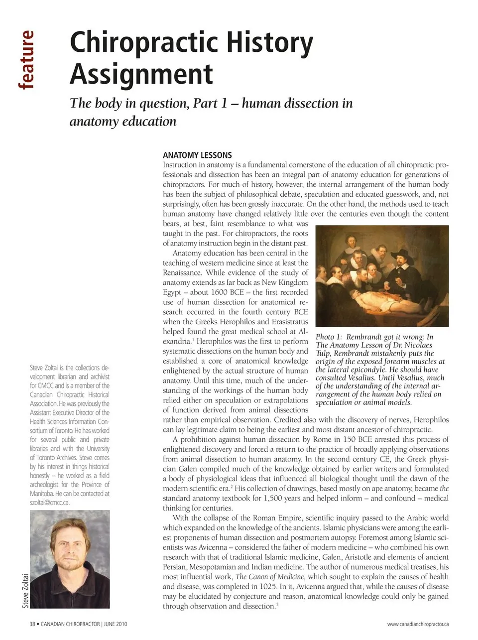
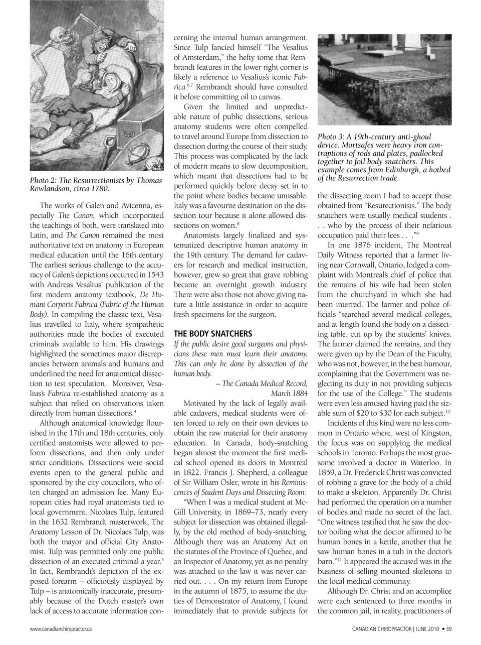
cerning the internal human arrangement. Since Tulp fancied himself “The Vesalius of Amsterdam,” the hefty tome that Rem- brandt features in the lower right corner is likely a reference to Vesalius’s iconic Fab- rica.6,7 Rembrandt should have consulted Photo 2: The Resurrectionists by Thomas Rowlandson, circa 1780. The works of Galen and Avicenna, es- pecially The Canon, which incorporated the teachings of both, were translated into Latin, and The Canon remained the most authoritative text on anatomy in European medical education until the 16th century. The earliest serious challenge to the accu- racy of Galen’s depictions occurred in 1543 with Andreas Vesalius’ publication of the first modern anatomy textbook, De Hu- mani Corporis Fabrica (Fabric of the Human Body). In compiling the classic text, Vesa- lius travelled to Italy, where sympathetic authorities made the bodies of executed criminals available to him. His drawings highlighted the sometimes major discrep- ancies between animals and humans and underlined the need for anatomical dissec- tion to test speculation. Moreover, Vesa- lius’s Fabrica re-established anatomy as a subject that relied on observations taken directly from human dissections.4 Although anatomical knowledge flour- ished in the 17th and 18th centuries, only certified anatomists were allowed to per- form dissections, and then only under strict conditions. Dissections were social events open to the general public and sponsored by the city councilors, who of- ten charged an admission fee. Many Eu- ropean cities had royal anatomists tied to local government. Nicolaes Tulp, featured in the 1632 Rembrandt masterwork, The Anatomy Lesson of Dr. Nicolaes Tulp, was both the mayor and official City Anato- mist. Tulp was permitted only one public dissection of an executed criminal a year.5 In fact, Rembrandt’s depiction of the ex- posed forearm – officiously displayed by Tulp – is anatomically inaccurate, presum- ably because of the Dutch master’s own lack of access to accurate information con- www.canadianchiropractor.ca it before committing oil to canvas. Given the limited and unpredict- able nature of public dissections, serious anatomy students were often compelled to travel around Europe from dissection to dissection during the course of their study. This process was complicated by the lack of modern means to slow decomposition, which meant that dissections had to be performed quickly before decay set in to the point where bodies became unusable. Italy was a favourite destination on the dis- section tour because it alone allowed dis- sections on women.8 Anatomists largely finalized and sys- tematized descriptive human anatomy in the 19th century. The demand for cadav- ers for research and medical instruction, however, grew so great that grave robbing became an overnight growth industry. There were also those not above giving na- ture a little assistance in order to acquire fresh specimens for the surgeon. THE BODY SNATCHERS If the public desire good surgeons and physi- cians these men must learn their anatomy. This can only be done by dissection of the human body. – The Canada Medical Record, March 1884 Motivated by the lack of legally avail- able cadavers, medical students were of- ten forced to rely on their own devices to obtain the raw material for their anatomy education. In Canada, body-snatching began almost the moment the first medi- cal school opened its doors in Montreal in 1822. Francis J. Shepherd, a colleague of Sir William Osler, wrote in his Reminis- cences of Student Days and Dissecting Room: “When I was a medical student at Mc- Gill University, in 1869–73, nearly every subject for dissection was obtained illegal- ly, by the old method of body-snatching. Although there was an Anatomy Act on the statutes of the Province of Quebec, and an Inspector of Anatomy, yet as no penalty was attached to the law it was never car- ried out. . . . On my return from Europe in the autumn of 1875, to assume the du- ties of Demonstrator of Anatomy, I found immediately that to provide subjects for Photo 3: A 19th-century anti-ghoul device. Mortsafes were heavy iron con- traptions of rods and plates, padlocked together to foil body snatchers. This example comes from Edinburgh, a hotbed of the Resurrection trade. the dissecting room I had to accept those obtained from “Resurectionists.” The body snatchers were usually medical students . . . who by the process of their nefarious occupation paid their fees . . .”9 In one 1876 incident, The Montreal It appeared the accused was in the business of selling mounted skeletons to the local medical community. Although Dr. Christ and an accomplice were each sentenced to three months in the common jail, in reality, practitioners of CANADIAN CHIROPRACTOR | JUNE 2010 • 39 Daily Witness reported that a farmer liv- ing near Cornwall, Ontario, lodged a com- plaint with Montreal’s chief of police that the remains of his wife had been stolen from the churchyard in which she had been interred. The farmer and police of- ficials “searched several medical colleges, and at length found the body on a dissect- ing table, cut up by the students’ knives. The farmer claimed the remains, and they were given up by the Dean of the Faculty, who was not, however, in the best humour, complaining that the Government was ne- glecting its duty in not providing subjects for the use of the College.” The students were even less amused having paid the siz- able sum of $20 to $30 for each subject.10 Incidents of this kind were no less com- mon in Ontario where, west of Kingston, the focus was on supplying the medical schools in Toronto. Perhaps the most grue- some involved a doctor in Waterloo. In 1859, a Dr. Frederick Christ was convicted of robbing a grave for the body of a child to make a skeleton. Apparently Dr. Christ had performed the operation on a number of bodies and made no secret of the fact. “One witness testified that he saw the doc- tor boiling what the doctor affirmed to be human bones in a kettle, another that he saw human bones in a tub in the doctor’s barn.”11
Chiropractic + Naturopathic Doctor June 2010: Page 39