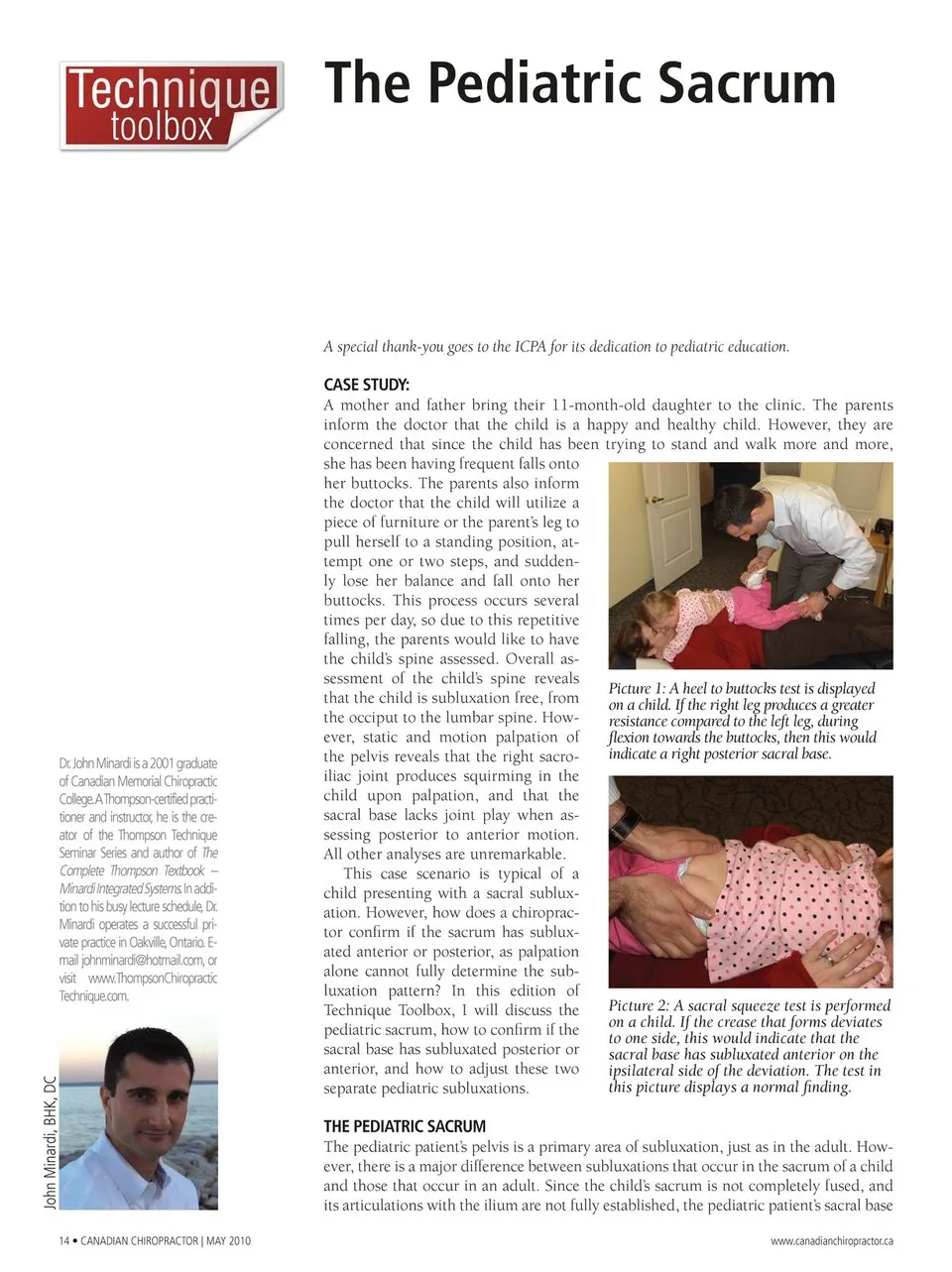
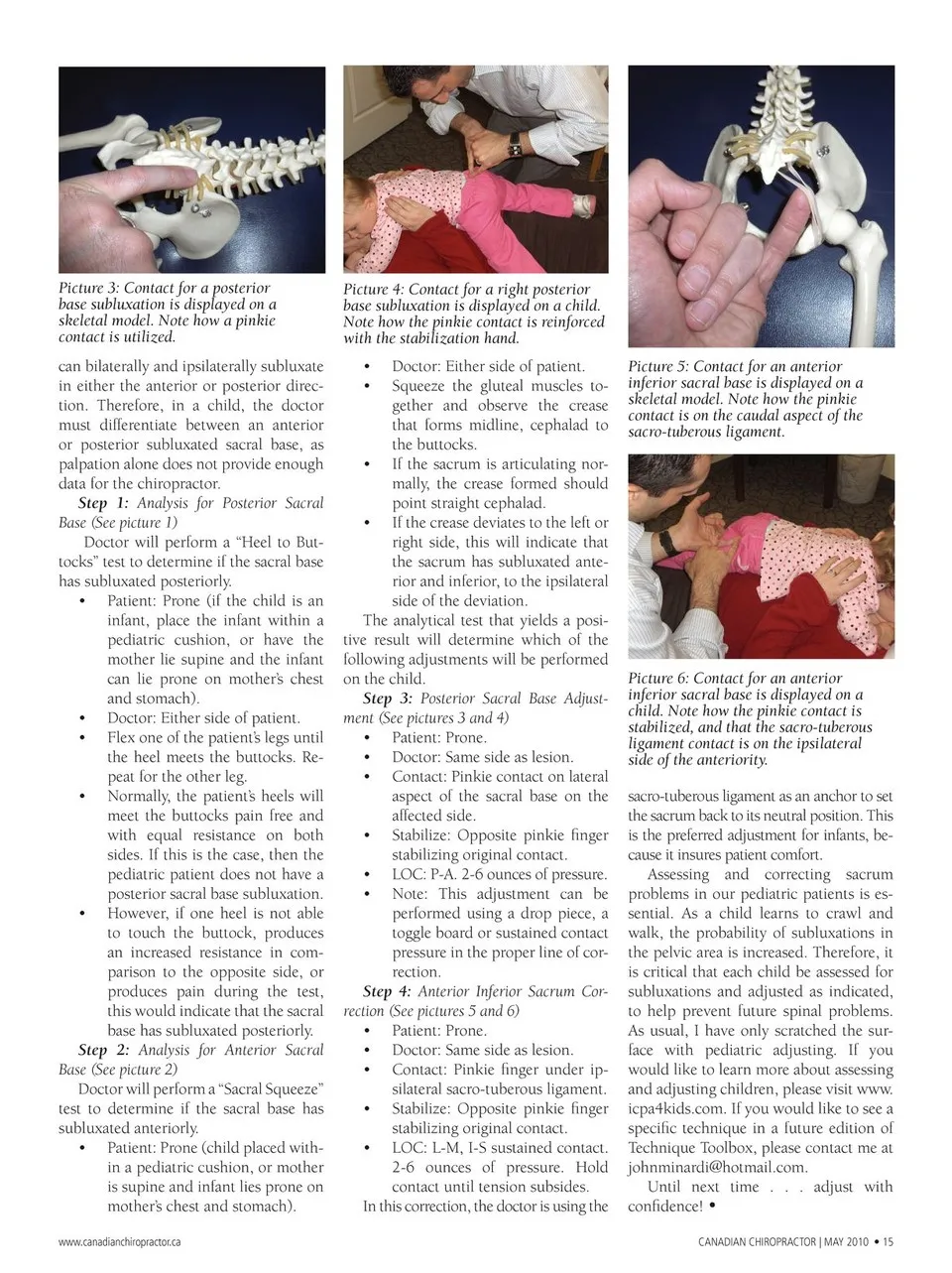
Picture 3: Contact for a posterior base subluxation is displayed on a skeletal model. Note how a pinkie contact is utilized. can bilaterally and ipsilaterally subluxate in either the anterior or posterior direc- tion. Therefore, in a child, the doctor must differentiate between an anterior or posterior subluxated sacral base, as palpation alone does not provide enough data for the chiropractor. Step 1: Analysis for Posterior Sacral Base (See picture 1) Doctor will perform a “Heel to But- tocks” test to determine if the sacral base has subluxated posteriorly. • Patient: Prone (if the child is an infant, place the infant within a pediatric cushion, or have the mother lie supine and the infant can lie prone on mother’s chest and stomach). • Doctor: Either side of patient. • Flex one of the patient’s legs until the heel meets the buttocks. Re- peat for the other leg. • Normally, the patient’s heels will meet the buttocks pain free and with equal resistance on both sides. If this is the case, then the pediatric patient does not have a posterior sacral base subluxation. • However, if one heel is not able to touch the buttock, produces an increased resistance in com- parison to the opposite side, or produces pain during the test, this would indicate that the sacral base has subluxated posteriorly. Step 2: Analysis for Anterior Sacral Base (See picture 2) Doctor will perform a “Sacral Squeeze” test to determine if the sacral base has subluxated anteriorly. • Patient: Prone (child placed with- in a pediatric cushion, or mother is supine and infant lies prone on mother’s chest and stomach). www.canadianchiropractor.ca Picture 4: Contact for a right posterior base subluxation is displayed on a child. Note how the pinkie contact is reinforced with the stabilization hand. • Doctor: Either side of patient. • Squeeze the gluteal muscles to- gether and observe the crease that forms midline, cephalad to the buttocks. • If the sacrum is articulating nor- mally, the crease formed should point straight cephalad. • If the crease deviates to the left or right side, this will indicate that the sacrum has subluxated ante- rior and inferior, to the ipsilateral side of the deviation. The analytical test that yields a posi- tive result will determine which of the following adjustments will be performed on the child. Step 3: Posterior Sacral Base Adjust- ment (See pictures 3 and 4) • Patient: Prone. • Doctor: Same side as lesion. • Contact: Pinkie contact on lateral aspect of the sacral base on the affected side. • Stabilize: Opposite pinkie finger stabilizing original contact. • LOC: P-A. 2-6 ounces of pressure. • Note: This adjustment can be performed using a drop piece, a toggle board or sustained contact pressure in the proper line of cor- rection. Step 4: Anterior Inferior Sacrum Cor- rection (See pictures 5 and 6) • Patient: Prone. • Doctor: Same side as lesion. • Contact: Pinkie finger under ip- silateral sacro-tuberous ligament. • Stabilize: Opposite pinkie finger stabilizing original contact. • LOC: L-M, I-S sustained contact. 2-6 ounces of pressure. Hold contact until tension subsides. In this correction, the doctor is using the Picture 6: Contact for an anterior inferior sacral base is displayed on a child. Note how the pinkie contact is stabilized, and that the sacro-tuberous ligament contact is on the ipsilateral side of the anteriority. sacro-tuberous ligament as an anchor to set the sacrum back to its neutral position. This is the preferred adjustment for infants, be- cause it insures patient comfort. Assessing and correcting sacrum problems in our pediatric patients is es- sential. As a child learns to crawl and walk, the probability of subluxations in the pelvic area is increased. Therefore, it is critical that each child be assessed for subluxations and adjusted as indicated, to help prevent future spinal problems. As usual, I have only scratched the sur- face with pediatric adjusting. If you would like to learn more about assessing and adjusting children, please visit www. icpa4kids.com. If you would like to see a specific technique in a future edition of Technique Toolbox, please contact me at [email protected]. Until next time . . . adjust with confidence! • CANADIAN CHIROPRACTOR | MAY 2010 • 15 Picture 5: Contact for an anterior inferior sacral base is displayed on a skeletal model. Note how the pinkie contact is on the caudal aspect of the sacro-tuberous ligament.
Chiropractic + Naturopathic Doctor May 2010: Page 15