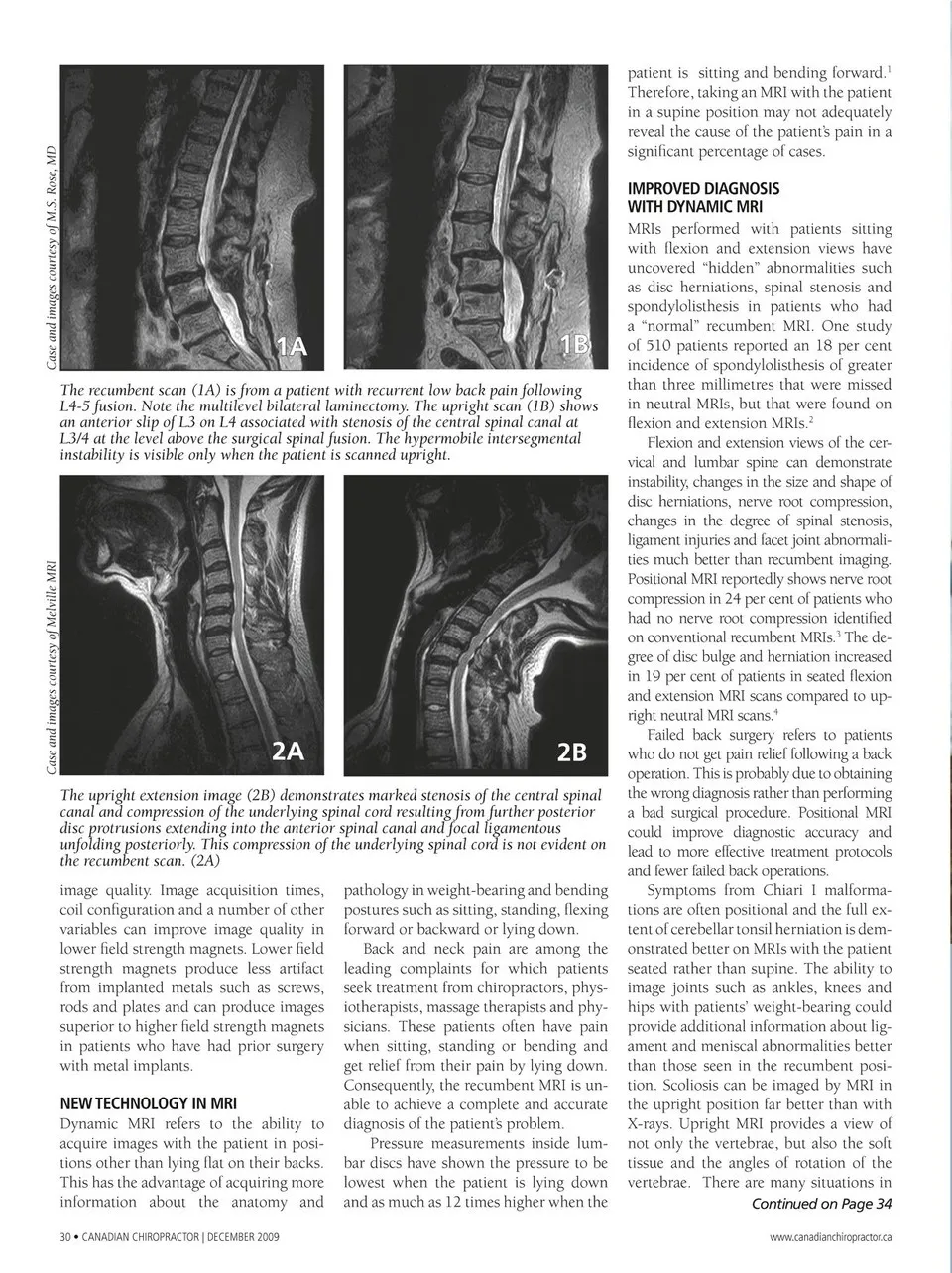
patient is sitting and bending forward.1 Therefore, taking an MRI with the patient in a supine position may not adequately reveal the cause of the patient’s pain in a significant percentage of cases. IMPROVED DIAGNOSIS WITH DYNAMIC MRI 1A 1B The recumbent scan (1A) is from a patient with recurrent low back pain following L4-5 fusion. Note the multilevel bilateral laminectomy. The upright scan (1B) shows an anterior slip of L3 on L4 associated with stenosis of the central spinal canal at L3/4 at the level above the surgical spinal fusion. The hypermobile intersegmental instability is visible only when the patient is scanned upright. MRIs performed with patients sitting with flexion and extension views have uncovered “hidden” abnormalities such as disc herniations, spinal stenosis and spondylolisthesis in patients who had a “normal” recumbent MRI. One study of 510 patients reported an 18 per cent incidence of spondylolisthesis of greater than three millimetres that were missed in neutral MRIs, but that were found on flexion and extension MRIs.2 Flexion and extension views of the cer- vical and lumbar spine can demonstrate instability, changes in the size and shape of disc herniations, nerve root compression, changes in the degree of spinal stenosis, ligament injuries and facet joint abnormali- ties much better than recumbent imaging. Positional MRI reportedly shows nerve root compression in 24 per cent of patients who had no nerve root compression identified on conventional recumbent MRIs.3 The de- 2A 2B The upright extension image (2B) demonstrates marked stenosis of the central spinal canal and compression of the underlying spinal cord resulting from further posterior disc protrusions extending into the anterior spinal canal and focal ligamentous unfolding posteriorly. This compression of the underlying spinal cord is not evident on the recumbent scan. (2A) image quality. Image acquisition times, coil configuration and a number of other variables can improve image quality in lower field strength magnets. Lower field strength magnets produce less artifact from implanted metals such as screws, rods and plates and can produce images superior to higher field strength magnets in patients who have had prior surgery with metal implants. NEW TECHNOLOGY IN MRI Dynamic MRI refers to the ability to acquire images with the patient in posi- tions other than lying flat on their backs. This has the advantage of acquiring more information about the anatomy and 30 • Canadian ChiropraCtor | dECEMBEr 2009 pathology in weight-bearing and bending postures such as sitting, standing, flexing forward or backward or lying down. Back and neck pain are among the leading complaints for which patients seek treatment from chiropractors, phys- iotherapists, massage therapists and phy- sicians. These patients often have pain when sitting, standing or bending and get relief from their pain by lying down. Consequently, the recumbent MRI is un- able to achieve a complete and accurate diagnosis of the patient’s problem. Pressure measurements inside lum- bar discs have shown the pressure to be lowest when the patient is lying down and as much as 12 times higher when the gree of disc bulge and herniation increased in 19 per cent of patients in seated flexion and extension MRI scans compared to up- right neutral MRI scans.4 Failed back surgery refers to patients who do not get pain relief following a back operation. This is probably due to obtaining the wrong diagnosis rather than performing a bad surgical procedure. Positional MRI could improve diagnostic accuracy and lead to more effective treatment protocols and fewer failed back operations. Symptoms from Chiari I malforma- tions are often positional and the full ex- tent of cerebellar tonsil herniation is dem- onstrated better on MRIs with the patient seated rather than supine. The ability to image joints such as ankles, knees and hips with patients’ weight-bearing could provide additional information about lig- ament and meniscal abnormalities better than those seen in the recumbent posi- tion. Scoliosis can be imaged by MRI in the upright position far better than with X-rays. Upright MRI provides a view of not only the vertebrae, but also the soft tissue and the angles of rotation of the vertebrae. There are many situations in Continued on Page 34 www.canadianchiropractor.ca Case and images courtesy of Melville MRI Case and images courtesy of M.S. Rose, MD
Chiropractic + Naturopathic Doctor December 2009: Page 30