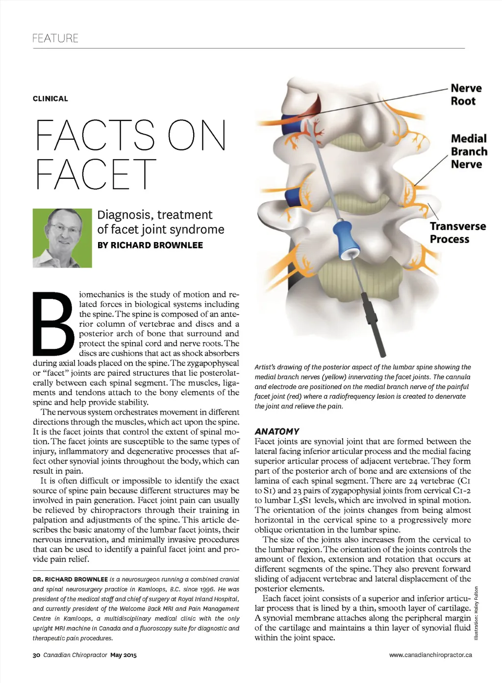
“Facet joint pain often occurs in episodic, severe exacerbations of pain that lasts for days.” Facet joints are endowed with intra-articular meniscoids that are composed of adipose tissue and blood vessels. They are located at the superior and inferior poles of the joint. The posterior surface of the joint is enclosed in a fibrous capsule of collagen fibers. The anterior surface is formed by the ligamentum flavum, which is a lateral ex-tension of the interlaminar ligament. pain. Therefore the source of a person’s pain cannot be determined solely by the morphologic appearance of the joints on imaging studies. FACET JOINT SYNDROME INNERVATION OF FACET JOINTS The lumbar facet joints are a recognized source of low back pain and referred leg pain. The term, facet syndrome was coined by Gormley in 1933. The prevalence of facet joint pain is reportedly between 15 and 40 per cent of people who suffer from chronic low back pain. Facet joints are a common source of neck pain following whiplash injury. While there is no pathogneumonic sign or symptom that distinguishes facet joint pain, it is generally accepted that facet joint pain is aggravated by extension and standing, as opposed to discogenic pain, which occurs with forward flexion or sitting. In addition to chronic back or neck pain, facet joint pain often occurs in episodic, severe exacerba-tions of pain that can last for days. People with facet joint pain often report collapsing or becoming completely immobilized by their pain. The sensory innervation to the facet joint is consistent. The spinal nerves at each level exit the intervertebral fo-ramen and divide into anterior and posterior primary rami. The posterior ramus, in turn, divides into a lateral branch that innervates the paraspinal muscles and a me-dial branch that provides sensation to the facet joints. The medial branch nerve runs across the top of the transverse process at its junction with the superior artic-ular process. Each facet joint receives sensory innervation from the medial branch nerve at the same vertebral level and from a descending branch from the level above. For example, the L4-5 facet joint is innervated by the L3 medial branch, which crosses the L4 transverse process and the L4 medial branch nerve, which crosses the L5 transverse process. The L5S1 facet joint is innervated by the L4 medial branch, which crosses the L5 transverse process and the L5 dorsal ramus, which crosses the Sacral Ala. To remove sensation from a painful facet joint, two medial branch nerves have to be anesthetized (blocked). The medial branch nerves also innervate the multifidus muscle, the interspinous muscle and interspinous liga-ment at the same segmental level as the named medial branch nerve. DIAGNOSTIC BLOCKS Determining if pain is arising from the facet joints and which joint is the source of the pain can be done through diagnostic medial branch blocks. This involves the injection -every major ideos V 190 muscle CHANGES WITH INJURY AND AGING Repeated stresses applied to the joints lead to progressive changes that may be classified as osteoarthrosis. This is not a disease but an expression of the morphologic con-sequence of stresses applied to the joints over time. Imaging studies with x-ray, CT and MRI often reveal changes, which include erosion and focal thinning of the cartilage, osteophyte formation at attachment sites of the capsular ligaments or ligamentum flavum, synovial cyst formation and enlargement of the joints. These changes occur with equal frequency in patients who have back or neck pain and in those who do not have pain. Patients who have normal appearing facet joints can also experience www.canadianchiropractor.ca The best app for patient education. Foam Roller Techniques puts a video library at their finger tips. Tap a muscle and watch the proper rolling technique demonstrated. Includes progression and regression variations as well as search by sport or injury. Based on the Roll Release® system by Dr. Ryan Emmons. FoamRollerApp.com May 2015 Canadian Chiropractor 31
Chiropractic + Naturopathic Doctor May 2015: Page 31