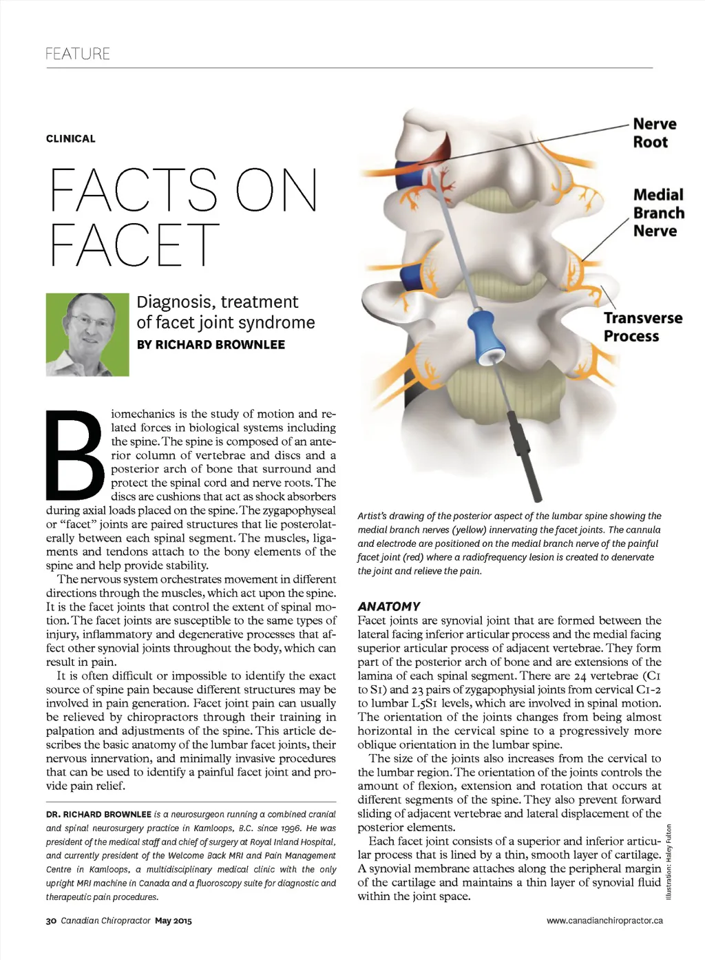
FEATURE CLINICAL FACTS ON FACET Diagnosis, treatment of facet joint syndrome BY RICHARD BROWNLEE B iomechanics is the study of motion and re-lated forces in biological systems including the spine. The spine is composed of an ante-rior column of vertebrae and discs and a posterior arch of bone that surround and protect the spinal cord and nerve roots. The discs are cushions that act as shock absorbers during axial loads placed on the spine. The zygapophyseal or “facet” joints are paired structures that lie posterolat-erally between each spinal segment. The muscles, liga-ments and tendons attach to the bony elements of the spine and help provide stability. The nervous system orchestrates movement in different directions through the muscles, which act upon the spine. It is the facet joints that control the extent of spinal mo-tion. The facet joints are susceptible to the same types of injury, inflammatory and degenerative processes that af-fect other synovial joints throughout the body, which can result in pain. It is often difficult or impossible to identify the exact source of spine pain because different structures may be involved in pain generation. Facet joint pain can usually be relieved by chiropractors through their training in palpation and adjustments of the spine. This article de-scribes the basic anatomy of the lumbar facet joints, their nervous innervation, and minimally invasive procedures that can be used to identify a painful facet joint and pro-vide pain relief. DR. RICHARD BROWNLEE is a neurosurgeon running a combined cranial and spinal neurosurgery practice in Kamloops, B.C. since 1996. He was president of the medical staff and chief of surgery at Royal Inland Hospital, and currently president of the Welcome Back MRI and Pain Management Centre in Kamloops, a multidisciplinary medical clinic with the only upright MRI machine in Canada and a fluoroscopy suite for diagnostic and therapeutic pain procedures. 30 Canadian Chiropractor May 2015 Artist’s drawing of the posterior aspect of the lumbar spine showing the medial branch nerves (yellow) innervating the facet joints. The cannula and electrode are positioned on the medial branch nerve of the painful facet joint (red) where a radiofrequency lesion is created to denervate the joint and relieve the pain. Facet joints are synovial joint that are formed between the lateral facing inferior articular process and the medial facing superior articular process of adjacent vertebrae. They form part of the posterior arch of bone and are extensions of the lamina of each spinal segment. There are 24 vertebrae (C1 to S1) and 23 pairs of zygapophysial joints from cervical C1-2 to lumbar L5S1 levels, which are involved in spinal motion. The orientation of the joints changes from being almost horizontal in the cervical spine to a progressively more oblique orientation in the lumbar spine. The size of the joints also increases from the cervical to the lumbar region. The orientation of the joints controls the amount of flexion, extension and rotation that occurs at different segments of the spine. They also prevent forward sliding of adjacent vertebrae and lateral displacement of the posterior elements. Each facet joint consists of a superior and inferior articu-lar process that is lined by a thin, smooth layer of cartilage. A synovial membrane attaches along the peripheral margin of the cartilage and maintains a thin layer of synovial fluid within the joint space. www.canadianchiropractor.ca ANATOMY Illustration: Haley Fulton
Chiropractic + Naturopathic Doctor May 2015: Page 30