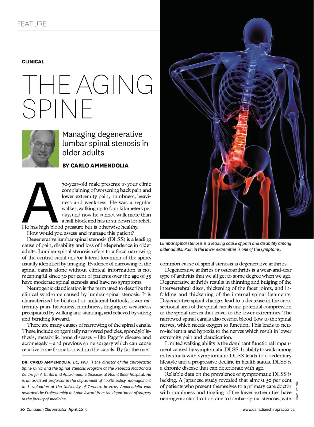
an average age of 65 years for females and 55 for males. An interesting phenomena associated with DLSS is the dynamic nature of the symptoms. Symptoms are usually precipitated by standing and walking. The longer one stands or walks the more intense the symptoms become. However, sitting or stooping forward or lying down results in a rapid reduction or elimination of the symptoms. This is explained by the change in the cross sectional area of spinal canals with changing posture. Lumbar flexion, which occurs when you sit or stoop forward, increases the spinal cross sectional area, reduces spinal nerve compression and restores spinal blood supply. Whereas, lumbar extension (we tend to maintain a lumbar lordosis when we stand and walk) decreases the cross sectional area and increases nerve pressure and symptoms. nerve compression. Loss of muscle tone and, in more ad-vanced cases, muscle atrophy are noted due to nerve com-pression and disuse (as a result of a more sedentary lifestyle). It is important to assess the lower extremity pulses to ensure they are present and symmetrically equal. DIFFERENTIAL DIAGNOSIS Inability to walk in patients with DLSS leads to a sedentary lifestyle and progressive decline in health. The patient described at the beginning of this article demonstrates this typical phenomenon and, at 70 years old, likely has degenerative changes in the spine. Patients with DLSS will typically describe their leg symptoms as numbness, tingling, pins and needles, weakness or heaviness in the but-tock, posterior thigh and lower leg that can impact their ability to walk. Back pain can be present, but not always, and can follow the same dynamic pattern. PHYSICAL EXAMINATION On physical examination, patients with DLSS tend to stand with a stooped posture. Range of motion testing typically demonstrates little difficulty during forward flexion. However, lumbar extension is usually limited and painful, and can sometimes reproduce lower extremity symptoms. Lumbar extension position may have to be maintained for a period of time before leg symptoms are reproduced. Balance testing may reveal difficulty and many patients will use a cane for added security. Heel-toe walking and a standing squat may demonstrate weakness that reflects involvement of a specific nerve root(s). However, this is usually seen in more long standing DLSS. The same holds true for sensory deficits, which will follow a dermatomal pattern. Deep tendon reflexes of the lower extremities are often difficult to elicit in older patients, although this is usually noted symmetrically. Supine straight leg raising is usually negative in DLSS be-cause this maneuver introduces lumbar flexion and reduces www.canadianchiropractor.ca One of the key goals in assessment is to determine the source of the patient’s symptoms and functional limitations in order to provide appropriate treatment. Osteoarthritis of the hip is highly prevalent in this patient population and can mimic DLSS. Symptoms are usually lo-cated in the back, buttock and groin, and pain can be referred distally to the knee. The symptoms are worse standing and walking and relieved by sitting and lying down. However, there tends to be a characteristic limping gait with a lateral shift in the pelvis on the involved side when weight bearing (Trendelenberg Sign) and usually no large change in symp-toms when walking with a stooped posture or when using a shopping cart. A hip exam is critical and often demonstrates painful and restricted internal rotation and flexion during range of motion testing. This testing usually reproduces the patient’s symptoms. Hip-spine syndrome is the diagnosis given when imaging confirmed osteoarthritis of the hip and DLSS are present at the same time, and both contribute to the patient’s symptoms and functional limitations. Vascular claudication is a condition that impacts walking ability and also highly present in older patients. It is usually a result of peripheral arterial disease (PVD) – the most common being due to atherosclerosis, which impacts blood flow to the lower extremities, especially the leg muscles. Like DLSS, symp-toms progress with walking and relieved by rest, but unlike DLSS stooped posture while walking does not change symp-toms or ability to walk. There can be trophic discolouration of skin noted in the lower extremities, especially involving the distal leg and foot. Diminished or absent pulses of the lower extremities can be noted but often not reliable for diagnosis. An arterial Doppler test is recommended to more accu-rately assess blood flow. About 30 per cent of DLSS patients also have confirmed PVD, which again makes diagnosis a challenge. Other conditions in the differential diagnosis of symptomatic DLSS include trochanteric bursitis, diabetic neuropathy and meralgia paresthetica. Disc herniation is more frequent in the 30 to 40 age groups and are usually associated with painful and restricted forward flexion, a positive straight leg raise and worsening symptoms with sitting, all of which are the antithesis of what is seen with DLSS. Neurogenic claudication is a clinical diagnosis based on the history and physical examination. Imaging is not necessary. Imaging is needed when red flags are present that suggest other potential serious diseases or conditions such as cancer, infection or spine fracture. Imaging is also needed when the patient’s condition is not improving and there may be the need for surgery or other invasive treatments. TREATMENT DLSS is the most common reason for spine surgery in individ-uals over the age of 65. There are generally two types of surgery: direct and indirect decompression. Direct decompression April 2015 Canadian Chiropractor 31
Chiropractic + Naturopathic Doctor April 2015: Page 31