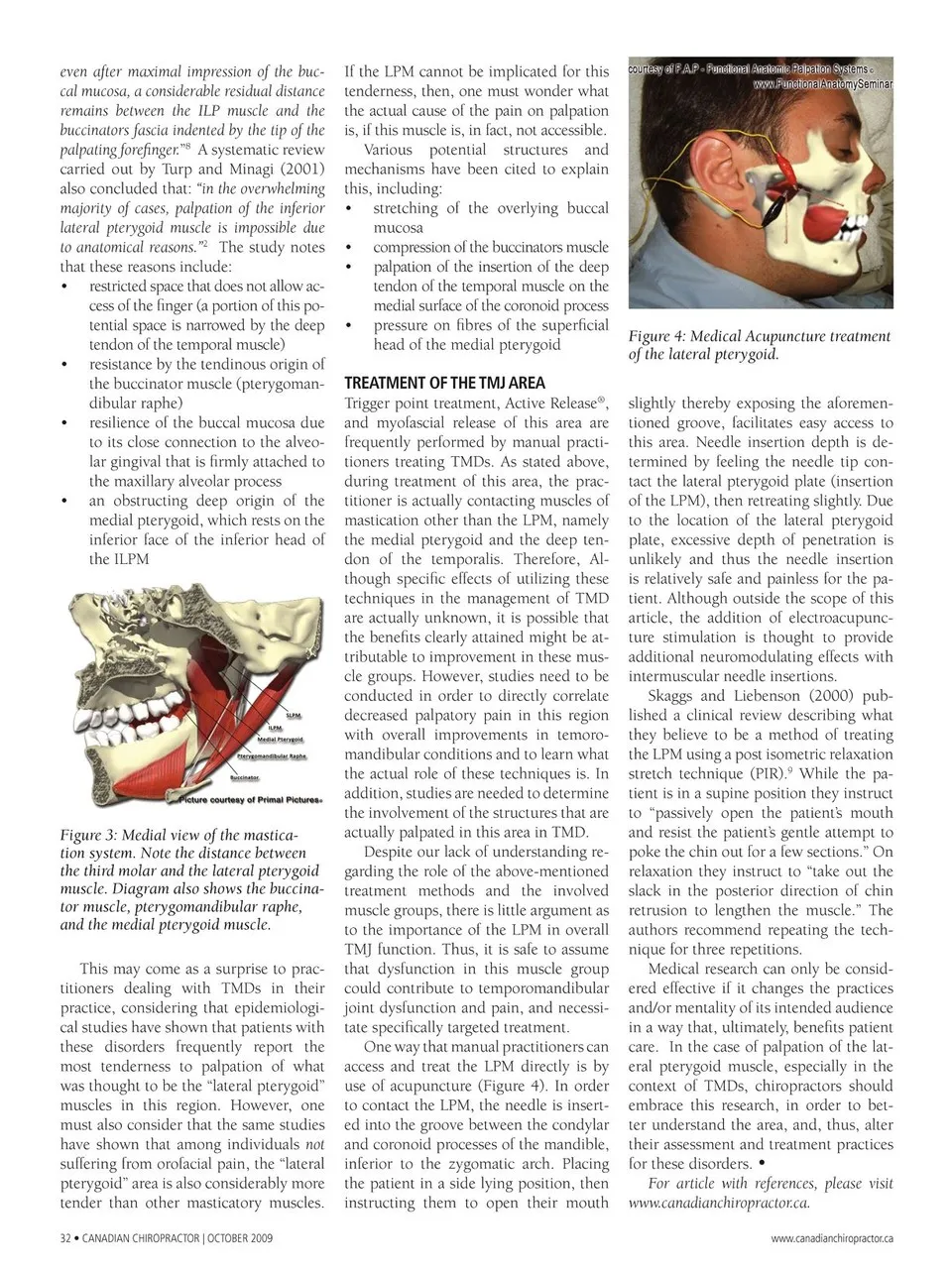
even after maximal impression of the buc- cal mucosa, a considerable residual distance remains between the ILP muscle and the buccinators fascia indented by the tip of the palpating forefinger.”8 A systematic review carried out by Turp and Minagi (2001) also concluded that: “in the overwhelming majority of cases, palpation of the inferior lateral pterygoid muscle is impossible due to anatomical reasons.”2 The study notes that these reasons include: restricted space that does not allow ac- cess of the fi nger (a portion of this po- tential space is narrowed by the deep tendon of the temporal muscle) resistance by the tendinous origin of the buccinator muscle (pterygoman- dibular raphe) resilience of the buccal mucosa due to its close connection to the alveo- lar gingival that is fi rmly attached to the maxillary alveolar process an obstructing deep origin of the medial pterygoid, which rests on the inferior face of the inferior head of the ILPM • • • • If the LPM cannot be implicated for this tenderness, then, one must wonder what the actual cause of the pain on palpation is, if this muscle is, in fact, not accessible. Various potential structures and mechanisms have been cited to explain this, including: stretching of the overlying buccal mucosa compression of the buccinators muscle palpation of the insertion of the deep tendon of the temporal muscle on the medial surface of the coronoid process pressure on fi bres of the superficial head of the medial pterygoid • • • • TREATMENT OF THE TMJ AREA Trigger point treatment, Active Release® , Figure 3: Medial view of the mastica- tion system. Note the distance between the third molar and the lateral pterygoid muscle. Diagram also shows the buccina- tor muscle, pterygomandibular raphe, and the medial pterygoid muscle. This may come as a surprise to prac- titioners dealing with TMDs in their practice, considering that epidemiologi- cal studies have shown that patients with these disorders frequently report the most tenderness to palpation of what was thought to be the “lateral pterygoid” muscles in this region. However, one must also consider that the same studies have shown that among individuals not suffering from orofacial pain, the “lateral pterygoid” area is also considerably more tender than other masticatory muscles. 32 • CANADIAN CHIROPRACTOR | OCTOBER 2009 and myofascial release of this area are frequently performed by manual practi- tioners treating TMDs. As stated above, during treatment of this area, the prac- titioner is actually contacting muscles of mastication other than the LPM, namely the medial pterygoid and the deep ten- don of the temporalis. Therefore, Al- though specifi c effects of utilizing these techniques in the management of TMD are actually unknown, it is possible that the benefi ts clearly attained might be at- tributable to improvement in these mus- cle groups. However, studies need to be conducted in order to directly correlate decreased palpatory pain in this region with overall improvements in temoro- mandibular conditions and to learn what the actual role of these techniques is. In addition, studies are needed to determine the involvement of the structures that are actually palpated in this area in TMD. Despite our lack of understanding re- garding the role of the above-mentioned treatment methods and the involved muscle groups, there is little argument as to the importance of the LPM in overall TMJ function. Thus, it is safe to assume that dysfunction in this muscle group could contribute to temporomandibular joint dysfunction and pain, and necessi- tate specifi cally targeted treatment. One way that manual practitioners can access and treat the LPM directly is by use of acupuncture (Figure 4). In order to contact the LPM, the needle is insert- ed into the groove between the condylar and coronoid processes of the mandible, inferior to the zygomatic arch. Placing the patient in a side lying position, then instructing them to open their mouth Figure 4: Medical Acupuncture treatment of the lateral pterygoid. slightly thereby exposing the aforemen- tioned groove, facilitates easy access to this area. Needle insertion depth is de- termined by feeling the needle tip con- tact the lateral pterygoid plate (insertion of the LPM), then retreating slightly. Due to the location of the lateral pterygoid plate, excessive depth of penetration is unlikely and thus the needle insertion is relatively safe and painless for the pa- tient. Although outside the scope of this article, the addition of electroacupunc- ture stimulation is thought to provide additional neuromodulating effects with intermuscular needle insertions. Skaggs and Liebenson (2000) pub- lished a clinical review describing what they believe to be a method of treating the LPM using a post isometric relaxation stretch technique (PIR).9 While the pa- tient is in a supine position they instruct to “passively open the patient’s mouth and resist the patient’s gentle attempt to poke the chin out for a few sections.” On relaxation they instruct to “take out the slack in the posterior direction of chin retrusion to lengthen the muscle.” The authors recommend repeating the tech- nique for three repetitions. Medical research can only be consid- ered effective if it changes the practices and/or mentality of its intended audience in a way that, ultimately, benefits patient care. In the case of palpation of the lat- eral pterygoid muscle, especially in the context of TMDs, chiropractors should embrace this research, in order to bet- ter understand the area, and, thus, alter their assessment and treatment practices for these disorders. • For article with references, please visit www.canadianchiropractor.ca. www.canadianchiropractor.ca
Chiropractic + Naturopathic Doctor October 2009: Page 32