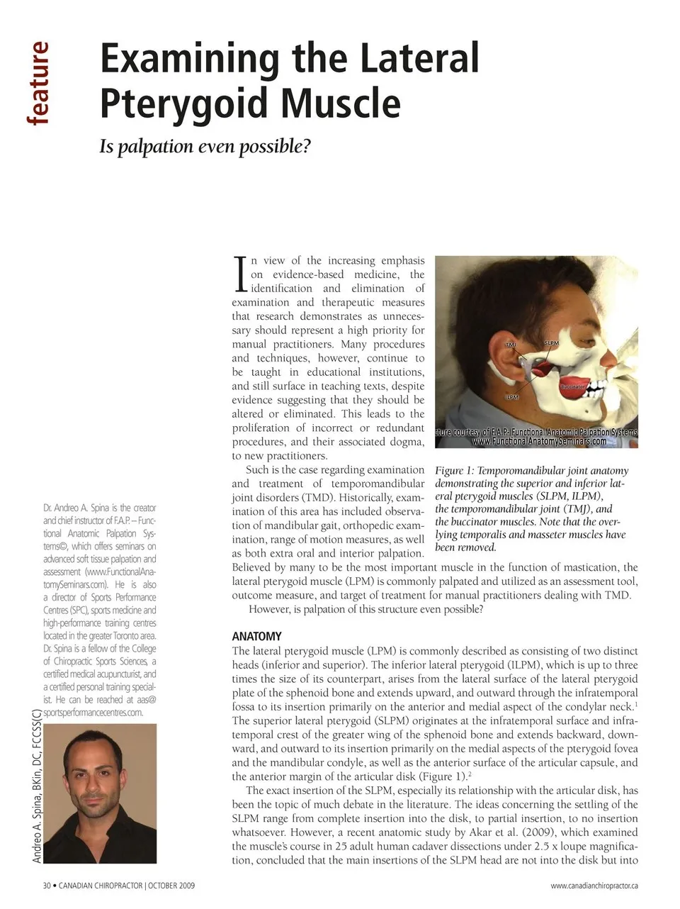
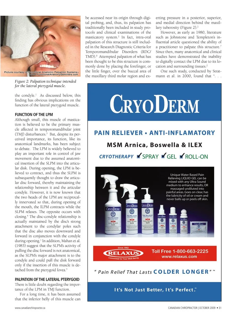
be accessed near its origin through digi- tal probing, and, thus, its palpation has traditionally been included in study pro- tocols and clinical examinations of the masticatory system.2 In fact, intra-oral Attempted palpation of what has been thought to be this structure is com- monly done by placing the forefinger, or the little fi nger, over the buccal area of the maxillary third molar region and ex- palpation of this structure is still includ- ed in the Research Diagnostic Criteria for Temporomandibular Disorders (RDC/ TMD).6 Figure 2: Palpation technique intended for the lateral pterygoid muscle. the condyle.1 As discussed below, this fi nding has obvious implications on the function of the lateral pterygoid muscle. FUNCTION OF THE LPM Although small, this muscle of mastica- tion is believed to be the primary mus- cle affected in temporomandibular joint (TMJ) disturbances.3 But, despite its per- The disc-condyle relationship is actually maintained by the disc’s strong attachment to the condylar poles such that the disc also moves downward and forward in conjunction with the condyle during opening.1 In addition, Mahan et al. (1983) suggest that the SLPM’s activity of pulling the disc forward is not anatomical, as the SLPM’s major attachment is to the condyle and could pull the disk forward only if the insertion of this muscle is de- tached from the pterygoid fovea.5 PALPATION OF THE LATERAL PTERYGOID There is little doubt regarding the impor- tance of the LPM in TMJ function. For a long time, it has been assumed that the inferior belly of this muscle can www.canadianchiropractor.ca CANADIAN CHIROPRACTOR | OCTOBER 2009 • 31 ceived importance, its function, like its anatomical landmarks, has been subject to debate. The LPM is widely believed to play an important role in control of jaw movement due to the assumed anatomi- cal insertion of the SLPM into the articu- lar disk. During opening, the LPM is be- lieved to contract, and thus the SLPM is subsequently thought to draw the articu- lar disc forward, thereby maintaining the relationship between it and the articular condyle. However, it is now known that the two heads of the LPM are reciprocal- ly innervated so that, during opening of the mouth, the ILPM contracts while the SLPM relaxes. The opposite occurs with closing.4 erting pressure in a posterior, superior, and medial direction behind the maxil- lary tuberosity (Figure 2).2 However, as early as 1980, literature such as Johnstone and Templeton’s in- fl uential article questioned the ability of a practitioner to palpate this structure.7 Since then, many anatomical and clinical studies have demonstrated the inability to digitally contact the LPM due to its lo- cation and surrounding tissues.2 One such study, conducted by Strat- mann et al. in 2000, found that “. . .
Chiropractic + Naturopathic Doctor October 2009: Page 31