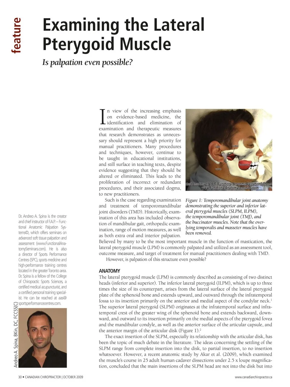
Examining the Lateral Pterygoid Muscle Is palpation even possible? I Dr. Andreo A. Spina is the creator and chief instructor of F.A.P.– Func- tional Anatomic Palpation Sys- tems©, which offers seminars on advanced soft tissue palpation and assessment (www.FunctionalAna- tomySeminars.com). He is also a director of Sports Performance Centres (SPC), sports medicine and high-performance training centres located in the greater Toronto area. Dr. Spina is a fellow of the College of Chiropractic Sports Sciences, a certified medical acupuncturist,and a certified personal training special- ist. He can be reached at aas@ sportsperformancecentres.com. n view of the increasing emphasis on evidence-based medicine, the identifi cation and elimination of examination and therapeutic measures that research demonstrates as unneces- sary should represent a high priority for manual practitioners. Many procedures and techniques, however, continue to be taught in educational institutions, and still surface in teaching texts, despite evidence suggesting that they should be altered or eliminated. This leads to the proliferation of incorrect or redundant procedures, and their associated dogma, to new practitioners. Such is the case regarding examination and treatment of temporomandibular joint disorders (TMD). Historically, exam- ination of this area has included observa- tion of mandibular gait, orthopedic exam- ination, range of motion measures, as well as both extra oral and interior palpation. Figure 1: Temporomandibular joint anatomy demonstrating the superior and inferior lat- eral pterygoid muscles (SLPM, ILPM), the temporomandibular joint (TMJ), and the buccinator muscles. Note that the over- lying temporalis and masseter muscles have been removed. Believed by many to be the most important muscle in the function of mastication, the lateral pterygoid muscle (LPM) is commonly palpated and utilized as an assessment tool, outcome measure, and target of treatment for manual practitioners dealing with TMD. However, is palpation of this structure even possible? ANATOMY The lateral pterygoid muscle (LPM) is commonly described as consisting of two distinct heads (inferior and superior). The inferior lateral pterygoid (ILPM), which is up to three times the size of its counterpart, arises from the lateral surface of the lateral pterygoid plate of the sphenoid bone and extends upward, and outward through the infratemporal fossa to its insertion primarily on the anterior and medial aspect of the condylar neck.1 The superior lateral pterygoid (SLPM) originates at the infratemporal surface and infra- temporal crest of the greater wing of the sphenoid bone and extends backward, down- ward, and outward to its insertion primarily on the medial aspects of the pterygoid fovea and the mandibular condyle, as well as the anterior surface of the articular capsule, and the anterior margin of the articular disk (Figure 1).2 The exact insertion of the SLPM, especially its relationship with the articular disk, has been the topic of much debate in the literature. The ideas concerning the settling of the SLPM range from complete insertion into the disk, to partial insertion, to no insertion whatsoever. However, a recent anatomic study by Akar et al. (2009), which examined the muscle’s course in 25 adult human cadaver dissections under 2.5 x loupe magnifica- tion, concluded that the main insertions of the SLPM head are not into the disk but into 30 • CANADIAN CHIROPRACTOR | OCTOBER 2009 www.canadianchiropractor.ca Andreo A. Spina, BKin, DC, FCCSS(C) feature
Chiropractic + Naturopathic Doctor October 2009: Page 30