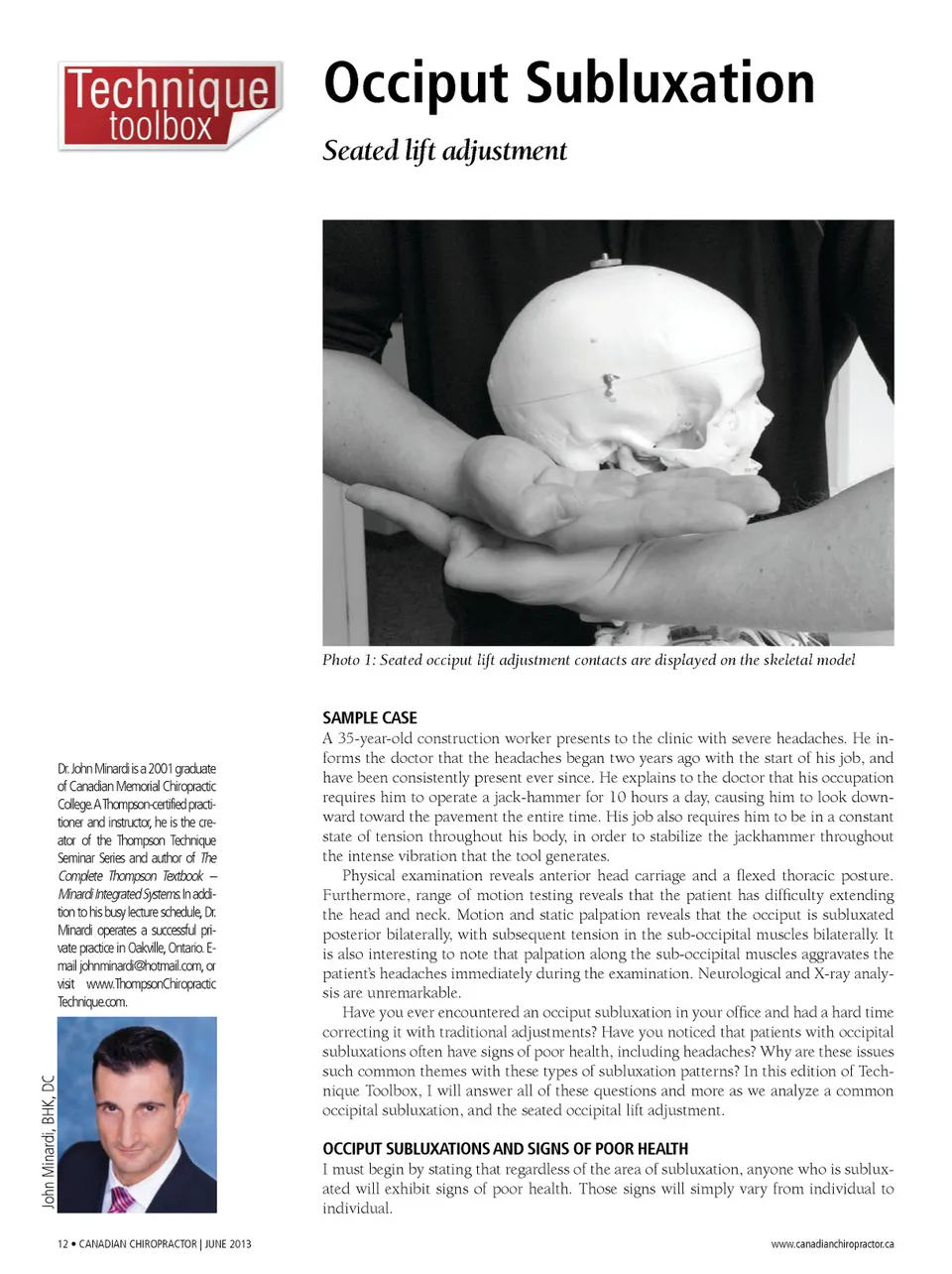
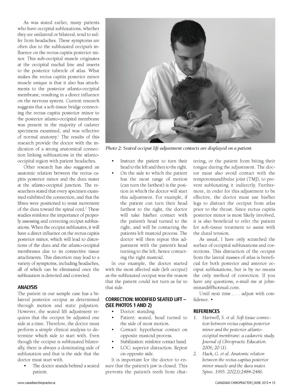
As was stated earlier, many patients who have occipital subluxations, whether they are unilateral or bilateral, tend to suf-fer from headaches. These symptoms are often due to the subluxated occiput’s in-fluence on the rectus capitis posterior mi-nor. This sub-occipital muscle originates at the occipital nuchal line and inserts to the posterior tubercle of atlas. What makes the rectus capitis posterior minor muscle unique is that it also has attach-ments to the posterior atlanto-occipital membrane, resulting in a direct influence on the nervous system. Current research suggests that a soft-tissue bridge connect-ing the rectus capitis posterior minor to the posterior atlanto-occipital membrane was present in the majority of cadaver specimens examined, and was reflective of normal anatomy. 1 The results of this research provide the doctor with the in-dication of a strong anatomical connec-tion linking subluxations in the atlanto-occipital region with patient headaches. Other research has also suggested an anatomic relation between the rectus ca-pitis posterior minor and the dura mater at the atlanto-occipital junction. The re-searchers stated that every specimen exam-ined exhibited the connection, and that the fibres were positioned to resist movement of the dura toward the spinal cord. 2 These studies reinforce the importance of proper-ly assessing and correcting occiput sublux-ations. When the occiput subluxates, it will have a direct influence on the rectus capitis posterior minor, which will lead to distor-tions of the dura and the atlanto-occipital membranes due to its connective tissue attachments. This distortion may lead to a variety of symptoms, including headaches, all of which can be eliminated once the subluxation is detected and corrected. Photo 2: Seated occiput lift adjustment contacts are displayed on a patient. Instruct the patient to turn their head to the left and then to the right. • On the side to which the patient has the most range of motion (can turn the farthest) is the posi-tion in which the doctor will start this adjustment. For example, if the patient can turn their head farthest to the right, the doctor will take his/her contact with the patient’s head turned to the right, and will be contacting the patients left mastoid process. The doctor will then repeat this ad-justment with the patient’s head turning to the left, hence contact-ing the right mastoid. In our example, the doctor started with the most affected side (left occiput) as the subluxated occiput was the reason that the patient could not turn as far to that side. • tering, or the patient from biting their tongue during the adjustment. The doc-tor must also avoid contact with the temporomandibular joint (TMJ), to pre-vent subluxating it indirectly. Further-more, in order for this adjustment to be effective, the doctor must use his/her legs to distract the occiput from atlas prior to the thrust. Since rectus capitis posterior minor is most likely involved, it is also beneficial to refer the patient for soft-tissue treatment to assist with the dural tension. As usual, I have only scratched the surface of occipital subluxations and cor-rections. This distraction of the occiput from the lateral masses of atlas is benefi-cial for both posterior and anterior oc-ciput subluxations, but is by no means the only method of correction. If you have any questions, e-mail me at [email protected]. Until next time . . . adjust with con-fidence. • ANALYSIS The patient in our sample case has a bi-lateral posterior occiput as determined through motion and static palpation. However, the seated lift adjustment re-quires that the occiput be adjusted one side at a time. Therefore, the doctor must perform a simple clinical analysis to de-termine which side to start with. Even though the occiput is subluxated bilater-ally, there is always a dominating side of subluxation and that is the side that the doctor must start with. • The doctor stands behind a seated patient. www.canadianchiropractor.ca CORRECTION: MODIFIED SEATED LIFT – (SEE PHOTOS 1 AND 2) • Doctor: standing. • Patient: seated, head turned to the side of most motion. • Contact: hypothenar contact on opposite mastoid process. • Stabilization: reinforce contact hand. • LOC: superior distraction. Repeat on opposite side. It is important for the doctor to en-sure that the patient’s jaw is closed. This prevents the patient’s teeth from chat-REFERENCES 1. Hartwell, S. et al. Soft tissue connec-tion between rectus capitus posterior minor and the posterior atlanto-occipital membrane: a cadaveric study. Journal of Chiropractic Education. 2006; 20 (1). 2. Hack, G. et al. Anatomic relation between the rectus capitus posterior minor muscle and the dura mater. Spine. 1995. 20(23):2484-2486. CANADIAN CHIROPRACTOR | JUNE 2013 • 13
Chiropractic + Naturopathic Doctor June 2013 : Page 13