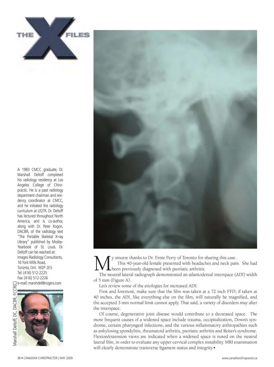
A 1983 CMCC graduate, Dr. Marshall Deltoff completed his radiology residency at Los Angeles College of Chiro- practic. He is a past radiology department chairman and resi- dency coordinator at CMCC, and he initiated the radiology curriculum at UQTR. Dr. Deltoff has lectured throughout North America, and is co-author, along with Dr. Peter Kogon, DACBR, of the radiology text “The Portable Skeletal X-ray Library” published by Mosby- Yearbook of St. Louis. Deltoff can be reached at: Images Radiology Consultants, 16 York Mills Road, Toronto, Ont. M2P 2E5 Tel: (416) 512-2225 Fax: (416) 512-2226 e-mail: [email protected] Dr. M 38 • CANADIAN CHIROPRACTOR | MAY 2009 y sincere thanks to Dr. Ernie Perry of Toronto for sharing this case. This 40-year-old female presented with headaches and neck pain. She had been previously diagnosed with psoriatic arthritis. The neutral lateral radiograph demonstrated an atlantodental interspace (ADI) width of 5 mm (Figure A). Let’s review some of the etiologies for increased ADI: First and foremost, make sure that the fi lm was taken at a 72 inch FFD; if taken at 40 inches, the ADI, like everything else on the fi lm, will naturally be magnified, and the accepted 3 mm normal limit cannot apply. That said, a variety of disorders may alter the interspace. Of course, degenerative joint disease would contribute to a decreased space. The more frequent causes of a widened space include trauma, occipitalization, Down’s syn- drome, certain pharyngeal infections, and the various infl ammatory arthropathies such as ankylosing spondylitis, rheumatoid arthritis, psoriatic arthritis and Reiter’s syndrome. Flexion/extension views are indicated when a widened space is noted on the neutral lateral fi lm, in order to evaluate any upper cervical complex instability. MRI examination will clearly demonstrate transverse ligament status and integrity.• www.canadianchiropractor.ca Marshall Deltoff, DC, DACBR, FCCR(C)
Chiropractic + Naturopathic Doctor May 2009: Page 38