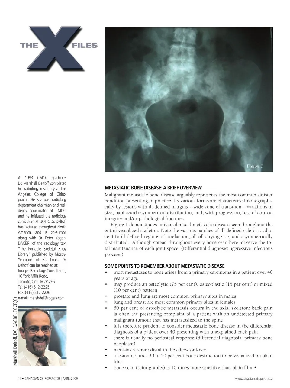
Figure 1 A 1983 CMCC graduate, Dr. Marshall Deltoff completed his radiology residency at Los Angeles College of Chiro- practic. He is a past radiology department chairman and resi- dency coordinator at CMCC, and he initiated the radiology curriculum at UQTR. Dr. Deltoff has lectured throughout North America, and is co-author, along with Dr. Peter Kogon, DACBR, of the radiology text “The Portable Skeletal X-ray Library” published by Mosby- Yearbook of St. Louis. Dr. Deltoff can be reached at: Images Radiology Consultants, 16 York Mills Road, Toronto, Ont. M2P 2E5 Tel: (416) 512-2225 Fax: (416) 512-2226 e-mail: [email protected] METASTATIC BONE DISEASE: A BrIEf OVErVIEW Malignant metastatic bone disease arguably represents the most common sinister condition presenting in practice. Its various forms are characterized radiographi- cally by lesions with ill-defined margins – wide zone of transition – variations in size, haphazard asymmetrical distribution, and, with progression, loss of cortical integrity and/or pathological fractures. Figure 1 demonstrates universal mixed metastatic disease seen throughout the entire visualized skeleton. Note the various patches of ill-defined sclerosis adja- cent to ill-defined regions of rarefaction, all of varying size, and asymmetrically distributed. Although spread throughout every bone seen here, observe the to- tal maintenance of each joint space. (Differential diagnosis: aggressive infectious process.) SOME POINTS TO rEMEMBEr ABOUT METASTATIC DISEASE • most metastases to bone arises from a primary carcinoma in a patient over 40 years of age • may produce an osteolytic (75 per cent), osteoblastic (15 per cent) or mixed (10 per cent) pattern • prostate and lung are most common primary sites in males • lung and breast are most common primary sites in females • 80 per cent of osteolytic metastasis occurs in the axial skeleton: back pain is often the presenting complaint of a patient with an undetected primary malignant tumour that has metastasized to the spine • it is therefore prudent to consider metastatic bone disease in the differential diagnosis of a patient over 40 presenting with unexplained back pain • there is usually no periosteal response (differential diagnosis: primary bone neoplasm) • metastasis is rare distal to the elbow or knee • a lesion requires 30 to 50 per cent bone destruction to be visualized on plain film • bone scan (scintigraphy) is 10 times more sensitive than plain film • 46 • CANADIAN CHIROPRACTOR | APRIL 2009 www.canadianchiropractor.ca Marshall Deltoff, DC, DACBR, FCCR(C)
Chiropractic + Naturopathic Doctor April 2009: Page 46